
Volume 20 Issue 2, February 2023
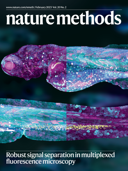
Robust signal separation in multiplexed fluorescence microscopy
Highly multiplexed fluorescence images of embryonic zebrafish using data analyzed with the Hybrid Unmixing (HyU) method.
See Chiang et al.
Image: H. J. Chiang, D. E. S. Koo, F. Cutrale, University of Southern California. Cover Design: Thomas Phillips.
Editorial
-
Advertisement
This Month
Correspondence
Comment
Research Highlights
Technology Feature
-
Actin in action
Collection:
