Thank you for visiting nature.com. You are using a browser version with limited support for CSS. To obtain
the best experience, we recommend you use a more up to date browser (or turn off compatibility mode in
Internet Explorer). In the meantime, to ensure continued support, we are displaying the site without styles
and JavaScript.
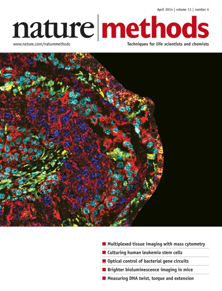
On the cover: Highly multiplexed analysis of human breast cancer tissue by imaging mass cytometry at 1-micrometer resolution. Image by Nicole Seidel, Graphic Design Seidel & Risse. Article p417
Nature Methods is dedicated to publishing methodological developments for basic biological research. Yet many papers that we receive, and some that we publish, also have later-stage applications. Where do we draw the line of editorial scope?
From single-molecule functional studies to atomic-resolution structures, a windfall of data sheds light on the Cas9 mechanism of targeted DNA scission.
A new device for injecting membrane-protein microcrystals into the path of an X-ray free-electron laser beam improves the efficiency of serial femtosecond crystallography.
By pushing throughput, single-cell transcript profiling can replace marker-based sorting and bulk RNA sequencing to redefine tissues from the bottom up.
The combination of mass spectroscopy with immunohistochemistry allows highly multiplexed, directly quantitative imaging of tissue samples for both basic and clinical research.
A comparative analysis of methods for scoring human sleep data, in particular sleep spindles, from encephalographic recordings is reported. The authors develop methods for crowdsourcing the identification of sleep spindles and compare the detection performance of experts, non-experts and automated algorithms.
The synthetic firefly luciferase substrate CycLuc1 offers brighter bioluminescence and improved imaging in mouse models at lower doses than the standard D-luciferin.
Four types of mass spectrometry (MS) data—label-free quantitative bottom-up proteomics, native MS, ion mobility–MS and cross-linking MS—are used to derive restraints for structural modeling of large protein assemblies.
Multivariate linear mixed models implemented in the GEMMA software package add speed, power and the ability to test for genome-wide associations between genetic polymorphisms and multiple correlated phenotypes.
The combination of stimulated Raman scattering (SRS) microscopy with an alkyne group as a Raman-active tag allows high-contrast, live-cell dynamic imaging of a variety of biomolecules including DNA, RNA, protein, lipids and small-molecule drugs.
CS-Rosetta-RNA is a web tool for determining high-resolution structures of noncoding RNA motifs using 1H NMR chemical shift data coupled with Rosetta-based modeling.
Cryo-scanning transmission electron tomography (CSTET) of unstained, fully hydrated vitrified biological specimens is shown to have advantages over cryo-electron tomography (CET), notably at high sample tilts providing greater depth resolution for thick samples.
High-throughput in vitro analysis of TALE nuclease (TALEN) cleavage specificity gives insight into the mechanism of TALE binding and allows the design of TALENs with lower off-target activity.
This paper reports conditions for the in vitro culture of human leukemia stem cells, which should enable basic studies as well as chemical screens on these cells.
Single-molecule structural transitions involving DNA twisting can be measured with substantially greater spatiotemporal resolution than previously possible with a gold rotor bead tracking (AuRBT) method. This approach uses magnetic tweezers and evanescent darkfield microscopy to track a gold nanoparticle probe attached to a DNA molecule.