Thank you for visiting nature.com. You are using a browser version with limited support for CSS. To obtain
the best experience, we recommend you use a more up to date browser (or turn off compatibility mode in
Internet Explorer). In the meantime, to ensure continued support, we are displaying the site without styles
and JavaScript.
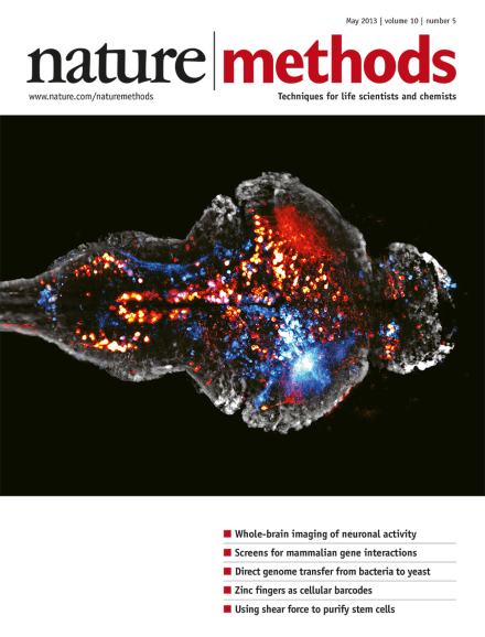
Bursts of neural activity in an entire larval zebrafish brain expressing a calcium reporter in every neuron. Two different time points are shown in red and blue. Image by Philipp Keller, Misha Ahrens and Kristin Branson (Howard Hughes Medical Institute, Janelia Farm Research Campus).
To produce enough raw material for high-quality whole-genome sequencing, researchers capture and grow single, culture-resistant bacteria in gel micro-droplets.
Zinc-finger DNA-binding domains are expressed on the surface of cells so that the cells can be tagged with fluorescently labeled oligonucleotides, enabling many possible applications in complex cell populations.
Surface expression of zinc-finger proteins coupled with double-stranded DNA probes permits programmable labeling and capture of subsets of cells in heterogenous populations.
Whole-brain imaging of neuronal activity with cellular resolution at almost a brain per second is demonstrated using high-speed light-sheet microscopy in the larval zebrafish brain.
Light-sheet microscopy using a laser beam reflected off a mirrored AFM cantilever provides high signal-to-background images suitable for high-speed quantitative single-molecule imaging of transcription factor binding to DNA in the nucleus of living mammalian cells.
Pairwise siRNA combinations and multiparametric imaging reveal positive and negative genetic interactions of epigenetic regulators in human cancer cells.
Differences in substrate adhesion strength are exploited in a label-free approach to separate human pluripotent stem cells from other cell types. This approach can separate fully and partially reprogrammed human induced pluripotent stem cells.