Thank you for visiting nature.com. You are using a browser version with limited support for CSS. To obtain
the best experience, we recommend you use a more up to date browser (or turn off compatibility mode in
Internet Explorer). In the meantime, to ensure continued support, we are displaying the site without styles
and JavaScript.
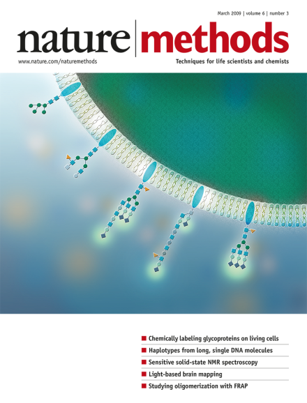
Sialic acidcontaining glycoproteins on mammalian cells can be tagged with fluorophores via an efficient chemical labeling approach. Cover design by Erin Boyle. Brief Communication, p207
The increasing impact of science on society calls for improved communication between scientists and the public via dedicated science media centers as well as nontraditional personal blogs.
A microscopy platform that brings magnetic resonance imaging to the nanometer scale offers a promising new tool for three-dimensional molecular visualization.
A method to improve the sensitivity of solid-state nuclear magnetic resonance spectroscopy promises to extend this technology to larger and more biologically interesting systems than previously feasible.
A map of single nucleotide polymorphisms (SNPs), also called a haplotype map, is very informative for mapping complex trait loci, but obtaining haplotypes over long genomic distances is very challenging. The combination of dye-labeling each SNP on PCR fragments with total internal reflection microscopy will allow the reading of long-range haplotypes with relative ease.
Absolute quantitative information about the stoichiometry of protein complex components can be obtained with a modified affinity purification–mass spectrometry method, as demonstrated for the human protein phosphatase 2A network.
Sialic acid–containing cell-surface glycoproteins can be chemically labeled with a biotin tag under mild conditions. The method is highly efficient and uses commercially available reagents; it should be useful for studying glycoprotein trafficking as well as in glycoproteomics applications.
As cells move over a substrate, they need to first sever their contact with the matrix by detaching focal adhesions. A setup that allows spatially and temporally controlled release of focal adhesions now facilitates the quantitative measurement of cell movement across a substrate.
Solid-state NMR spectroscopy is used to elucidate structural details about proteins that cannot be easily studied by X-ray crystallography, but because the technique is not very sensitive, large sample amounts are required, limiting its biological application. A combination of optimizations now increases the sensitivity of solid-state NMR spectroscopy by up to 5-fold.
Optical stimulation of channelrhodopsin-2 expressed in neurons of the motor cortex is combined with electromyogram recordings or motion-sensing of limb muscles to achieve fast motor mapping in the mouse.
Dual-color fluorescence recovery after photobleaching (FRAP) is used to investigate dimerization and higher-order complex formation of receptors at the surface of live cells. A defined fraction of receptors is immobilized with antibodies, and the mobility of the nonimmobilized fraction is measured by FRAP.
Small RNA discovery and profiling efforts are dramatically reshaping fundamental concepts of how genes are regulated and are leading to new tools for studying gene function.