
Thank you for visiting nature.com. You are using a browser version with limited support for CSS. To obtain the best experience, we recommend you use a more up to date browser (or turn off compatibility mode in Internet Explorer). In the meantime, to ensure continued support, we are displaying the site without styles and JavaScript.

Volume 6 Issue 10, October 2009

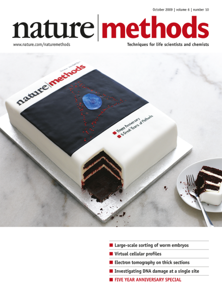
This delicious version of Nature Methods' inaugural issue was baked by How Sweet It Is (http://www.howsweetitispastry.com/), New York. Labeling of gangliosides in cellular membrane and nucleus is in red and blue fondant, respectively. Photography by Christina Holmes. Special 5th anniversary issue feature starts on p701.

Editorial

  • As evidenced by the cake adorning the cover, Nature Methods is five years old. To celebrate this anniversary, we look at methodological development and its role in scientific inquiry.

    Editorial

    Advertisement

Top of page ⤴

Correspondence

Top of page ⤴

Research Highlights

  • Channelrhodopsin-2 can be efficiently activated by infrared two-photon excitation light and stimulates action potentials in cultured neurons.

    • Natalie de Souza
    Research Highlights
  • By fusing a light-sensitive domain of an oat plant protein to Rac1, researchers created a genetically encoded protein fusion that can be reversibly activated with blue light and control cell movement—an attractive alternative to current caging tools.

    • Irene Kaganman
    Research Highlights
  • The development of leader sequences that stimulate mRNA translation in a species-independent manner could offer new possibilities for eukaryotic protein production and proteomic research.

    • Michael Eisenstein
    Research Highlights
  • Visualization of choline-containing phospholipids in cells and in vivo is made possible by the metabolic incorporation of a choline analog with an alkyne handle for click chemistry–based labeling.

    • Allison Doerr
    Research Highlights
  • Bacterial genomes shuttled into yeast can be easily altered before transplantation back into bacteria.

    • Nicole Rusk
    Research Highlights
Top of page ⤴

Historical Commentary

  • From histology to microcinematography, from cytochemistry to live cell imaging, the history of visualization technology in the life sciences may be understood as a series of cycles of action and reaction between static and dynamic modes of representing life.

    • Hannah Landecker
    Historical Commentary
Top of page ⤴

Commentary

  • Despite expansion of the fluorescent protein and optical highlighter palette into the orange to far-red range of the visible spectrum, achieving performance equivalent to that of EGFP has continued to elude protein engineers.

    • Michael W Davidson
    • Robert E Campbell
    Commentary
  • The potential of mass spectrometry–based proteomics to advance biology and biomedicine is nearly unlimited but so is its potential for generating bad data. Apart from the pursuit of technological progress in protocols and instruments, stringent comparative analyses of different approaches are critical for fully developing the discipline.

    • Matthias Mann
    Commentary
Top of page ⤴

News & Views

  • Automated stage-specific sorting of Caenorhabditis elegans embryos enables next-generation molecular and biochemical analysis of development.

    • L Ryan Baugh
    News & Views
Top of page ⤴

Brief Communication

  • Using an axial detector, scanning transmission electron microscopy allows three-dimensional tomographic reconstruction of micrometer-thick sections of biological samples, at a resolution comparable to that obtained on thin sections.

    • Martin F Hohmann-Marriott
    • Alioscka A Sousa
    • Richard D Leapman
    Brief Communication
  • Technical modifications of existing methods lead to a somatic cell nuclear transfer method in the zebrafish, which yields adult cloned fish with healthy offspring that carry donor traits.

    • Kannika Siripattarapravat
    • Boonya Pinmee
    • Jose B Cibelli
    Brief Communication
  • A series of genetically encoded fluorescent sensors for intracellular Zn2+ with binding affinities spanning the picomolar and nanomolar ranges show that cytosolic Zn2+ is buffered at 0.4 nM. Targeting of the sensors to insulin-containing secretory granules indicates a free Zn2+ concentration between 1 and 100 μM in these organelles.

    • Jan L Vinkenborg
    • Tamara J Nicolson
    • Maarten Merkx
    Brief Communication
  • Quantitative information is necessary to determine which protein interactions are the most relevant in a cellular context. A defined set of affinity purification experiments combined with quantitative mass spectrometry analysis allows the determination of dissociation constants for all protein targets interacting with an introduced ligand.

    • Kirti Sharma
    • Christoph Weber
    • Henrik Daub
    Brief Communication
Top of page ⤴

Article

Top of page ⤴

Technology Feature

  • Neuroscientists are taking advantage of powerful new tools for fluorescence imaging that enable detailed visualization of the structure and activity of neuronal circuits within the living brain.

    • Michael Eisenstein
    Technology Feature
Top of page ⤴

Advertising Feature: Application Note

Top of page ⤴

Focus

  • Nature Methodscelebrates its five year anniversary with commentaries discussing the impact and progress of methodological developments in the life sciences. We also include a fun selection of papers and covers from our pages.

    Focus
Top of page ⤴

Search

Quick links