Thank you for visiting nature.com. You are using a browser version with limited support for CSS. To obtain
the best experience, we recommend you use a more up to date browser (or turn off compatibility mode in
Internet Explorer). In the meantime, to ensure continued support, we are displaying the site without styles
and JavaScript.
Several research groups are making it easier for other neuroscientists to analyze large datasets by providing tools that can be accessed and used from anywhere in the world.
The exceptionally photostable green fluorescent protein StayGold has been monomerized in different laboratories, which has generated three unique monomeric variants that will enable new imaging applications.
New condenser aperture designs form square or rectangular beams that match the camera dimensions, which efficiently expands the data acquisition area in cryogenic electron microscopy.
Two studies show that nanopores can identify the 20 proteinogenic amino acids and some of their post-translational modifications. Coupled with an exopeptidase, a bottom-up approach to protein sequencing using nanopores is on the horizon.
How accurate is the prediction of protein structure by AlphaFold? Terwilliger et al. address this question with a rigorous assessment of the accuracy of AlphaFold-predicted structures by comparing them with experimentally determined X-ray crystallographic data.
A class of protein-based molecular shape probes move us closer toward the goal of a general, genetically encoded tagging system for cryogenic electron tomography.
Multiplexed spatial immunophenotyping has advanced our understanding of tissues in the context of homeostasis and disease. Two studies now provide additional tools to overcome challenges with multiplexed imaging: one procedure amplifies the detection of low-abundance antigens by integrating SABER and IMC technologies, and the other is an X-ray-based method that enables the non-destructive multiplexed detection of antigens in tissues at scalable resolution and speed.
A new chemically induced dimerization (CID) pair exhibits fluorescence upon dimerization for the first time. Moreover, the CID pair is small and offers easily reversible dimerization that can be repeated multiple times.
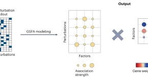
Recently proposed computational approaches explore casual links between chromatin and transcriptional changes that are provided by single-cell multimodal sequencing to bridge the knowledge gap in transcriptional regulatory control.
Three groundbreaking studies have created a new generation of genetically encoded voltage indicators, empowering us to tackle a host of questions on our path toward understanding the brain.
Nano-DMS-MaP focuses in on the structures of individual RNA isoforms, enabling direct examination of the structural diversity of different RNAs inside cells.
Two new Brillouin microscopes leverage line-scanning to overcome previous limitations of the technique, enabling fast imaging, with low phototoxicity, of mechanical properties in living embryos of model organisms and tumor spheroids.
Integration of single-cell molecular profiling with cellular spatial localization has remained an elusive goal. Image-seq leverages high-resolution microscopy to spatially resolve and isolate viable bone marrow and leukemia cells for subsequent state-of-the art, single-cell transcriptomics.