
Volume 20 Issue 4, April 2019
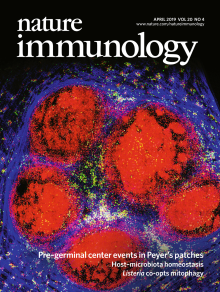
Pre-germinal center events in Peyer’s patches
Shulman and colleagues show that in Peyer’s patches, in contrast to lymph nodes and spleen, T cells predominantly promoted expansion of B cells without clonal selection during pre-germinal centre events.
See Shulman et al.
Image: Adi Biram. Cover Design: Erin Dewalt.
Research Highlights
-
Advertisement
