Thank you for visiting nature.com. You are using a browser version with limited support for CSS. To obtain
the best experience, we recommend you use a more up to date browser (or turn off compatibility mode in
Internet Explorer). In the meantime, to ensure continued support, we are displaying the site without styles
and JavaScript.
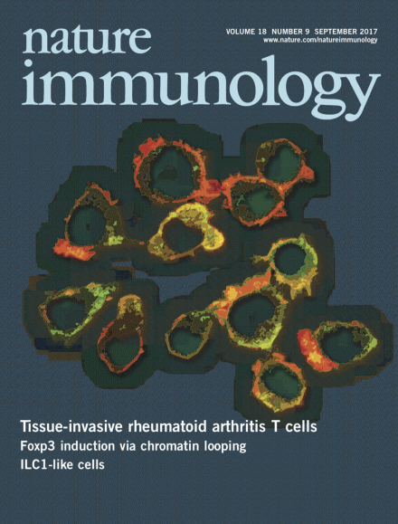
In rheumatoid arthritis, CD4+ T cells infiltration in joint tissues requires cytoskeletal reorganization and the formation of membrane protrusions. Shen and colleagues (p 1025; News and Views by Tsokos, p 955) show that CD4+ T cells from a person with rheumatoid arthritis are poised to form lamellopodia and membrane ruffles and be tissue invasive as a result of metabolic reprogramming. The original image by Yi Shen and Cornelia Weyand shows membrane ruffles and podosomes in CD4+ T cells from a person with rheumatoid arthritis. Artwork by Lewis Long.
The ability to expand and contract populations of myeloid and lymphoid cells during emergency hematopoiesis helps shape the immune response. The expression of intracellular and soluble forms of osteopontin regulates apoptosis thresholds differently in myeloid cells and lymphoid cells to counter infection.
Poor glycolysis and increased fatty-acid synthesis feed the locomotion machinery in T cells from people with rheumatoid arthritis and allow these cells to enter the synovium and propagate joint inflammation and destruction.
The histone lysine methyltransferase MLL4 primes the locus encoding the transcription factor Foxp3 for transcriptional activation in thymus-derived and inducible regulatory T cells.
The cytokine TGF-β allows tumors to evade the immune system by converting conventional natural killer cells into type 1 innate lymphoid cells devoid of cytotoxic function.
Long non-coding RNAs (lncRNAs) are being increasingly appreciated as important regulators of gene expression. Chang and colleagues review the roles identified for lncRNAs in the immune system and discuss models for how lncRNAs mediate their effects.
The size of immune-cell populations needs to be tightly regulated. Shinohara and colleagues demonstrate that intracellular and secreted osteopontin control the contraction and expansion of myeloid and lymphoid populations differently under infectious and autoimmune conditions.
How glutamine metabolism orchestrates macrophage activation is unclear. Ho and colleagues show glutamine metabolism tailors the immune responses of macrophages through metabolic and epigenetic reprogramming.
Colonna and colleagues generate mice in which both type 1 innate lymphoid cells and natural killer cells selectively lack SMAD4, which promotes canonical signaling of all cytokines of the TGF-β family. Unexpectedly, SMAD4 deficiency does not visibly affect the differentiation of type 1 innate lymphoid cells but instead alters the phenotype of conventional natural killer cells.
How tumors evade control by natural killer cells is ill defined. Smyth and colleagues show that natural killer cells in the tumor microenvironment can convert into type I innate lymphoid cells and intermediate type I innate lymphoid cells that favor tumor growth and metastasis.
Cathepsin S is responsible for cleaving the invariant chain Ii for the presentation of peptides by major histocompatibility complex class II on DCs. Diamond and colleagues show that the transcription factor Blimp-1 negatively regulates expression of cathepsin S and influences selection of the TFH cell repertoire and autoantibody-producing B cells.
Previous studies have defined a state of metabolic reprogramming in rheumatoid arthritis (RA) T cells. Here Weyand and colleagues show that the rewiring of cellular metabolism renders RA T cells tissue invasive, directly promoting disease-inducing effector functions.
Zhao and colleagues show that the histone lysine methyltransferase MLL4 primes the Foxp3 locus for transcriptional activation in thymus-derived and inducible regulatory T cells.
T cells undergo myriad changes after antigenic activation. Araki and colleagues show that CD8+ T cells exert dynamic control of mRNA translation during differentiation into effector and memory cells.