Volume 16
-
No. 12 December 2015
A safe mechanism for enhancing interferon signaling should improve immunity. Holtzman and colleagues increase interferon production by using a modified form of the transcription factor STAT1 to enhance expression of the PARP9-DTX3L ubiquitin ligase complex (p 1215). The original confocal image by Xiaohua Jin and Yong Zhang shows localization of DTX3L together with encephalomyocarditis virus 3C protease in the nucleus and cytosol of U3A human fibrosarcoma cells overexpressing DTX3L. Artwork by Lewis Long.
-
No. 11 November 2015
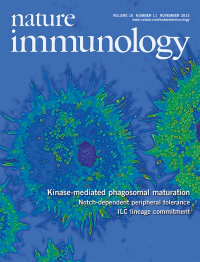
Reactive oxygen species (ROS) produced by phagocytes are critical for killing pathogens. Zhou and colleagues (p 1142; News and Views by Stuart and Lacy-Hulbert p 1107) show that the kinases Mst1 and Mst2 act through the small GTPase Rac to position mitochondria next to phagosomes and promote ROS production in phagocytes. The original image by Jing Geng, Lanfen Chen and Dawang Zhou shows bone marrowderived macrophages expressing a constitutively active form of Rac1, stained for F-actin (green) and for nuclei (with DAPI; blue). Artwork by Lewis Long.
-
No. 10 October 2015
Treatment with ionizing irradiation (IR) can lead to accumulation of tumor-infiltrating T regulatory (Treg) cells. Merad and colleagues (p 1060; News and Views by Zitvogel and Kroemer, p 1005) show that Langherans cells resist IR via expression of p21 and potentiate the generation and accumulation of Treg cells. The original image by Juliana Idoyaga shows immunofluorescence staining of a mouse epidermal sheet that reveals the presence of a dense cellular network of Langerhans cells stained with Langerin (CD207, green), MHC class II (red) and DAPI (blue). Artwork by Lewis Long.
-
No. 9 September 2015
Paneth cell dysfunction has been implicated in Crohn's disease. Liu and colleagues show that deficiency in the vesicle transport regulator LRRK2 leads to lysosomal degradation of lysozyme in Paneth cells (p 918; News and Views by Rocha, Schlossmacher and Philpott, p 898). The confocal image shows restored lysozyme staining (red) in cultured Lrrk2–/– intestinal organoids after lysosomal inhibition with leupeptin. Procryptin is stained green. Original image by Qin Zhang. Artwork by Lewis Long.
-
No. 8 August 2015
Fat-associated lymphoid clusters (FALCs) are a type of lymphoid tissue associated with visceral fat present in the mesenteries, mediastinum and pericardium. Benezech, Caamaño and colleagues show that FALCs support B cell proliferation and germinal center differentiation and that their formation is driven by inflammation (p 819; News and Views, p 796). The original image by Lucy Jones shows immunofluorescence staining of a mediastinal FALC with adipocytes (green), IgM+ B cells (red) and DAPI (blue). Artwork by Lewis Long.
-
No. 7 July 2015
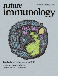
Antibody-secreting cells vastly increase in abundance during acute flares of systemic lupus erythematosus. Sanz and colleagues (p 755; News and Views by Tarlinton and Smith, p 685) show that this population results from polyclonal activation of B cells, including many derived from newly activated naive cells. The original image by Igor Albizua (Emory University) shows a transmission electron micrograph of a human antibody-secreting cell. Artwork by Lewis Long.
-
No. 6 June 2015
This month's Focus features a series of specially commissioned Reviews and a Perspective that discuss the most recent progress in understanding the immune response to HIV and how this new insight can be harnessed for prophylactic vaccines and immunotherapies (http://www.nature.com/ni/focus/hiv/index.html). Artwork by Lewis Long depicts an HIV-1 virion.
Focus
-
No. 5 May 2015
The histone methyltransferase Ezh2 contributes to epigenetic regulation. Su and colleagues show that Ezh2 also plays a critical cytosolic role by regulating leukocyte migration and adhesion dynamics via direct methylation of talin (p 505; News and Views by Wehrle-Haller, p 441). Original image by Nandini Venkatesan shows H16N2 epithelial cells expressing talin-GFP (green) and stained for F-actin (red) and nuclear DAPI (blue). Artwork by Lewis Long.
-
No. 4 April 2015
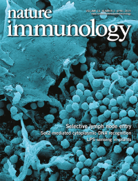
Lymph-borne lymphocytes and soluble antigens can traverse the lymphatic endothelial cells (LECs) lining the subcapsular sinus (SCS) of lymph nodes. Salmi and colleagues show that the endothelial protein Plvap forms a sieve which regulates these processes (p 386; and News and Views by Hons and Sixt, p 338). The image shows lymphocytes (round cells), SCS macrophages (cauliflower-like projections) and the floor LECs in a SCS of a mouse lymph node. Original image by Kazuo Tohya.Artwork by Lewis Long.
-
No. 3 March 2015
Intracellular bacteria have evolved sophisticated strategies to subvert the innate immune system, which allows them to survive inside macrophages for long periods. Liu and colleagues (p 237) demonstrate that Mycobacterium tuberculosis secretes the protein phosphatase PtpA, which is activated by host ubiquitin, to suppress innate immunity. Original image by Jing Wang and Cui Hua Liu shows a HeLa cell co-transfected with vectors encoding green fluorescent protein (green) and hemagglutinin-tagged ubiquitin (red).Artwork by Cameron Long and Lewis Long.
-
No. 2 February 2015
The deposition of antibody-antigen complexes in kidney glomeruli is a frequent occurrence in systemic autoimmune diseases. Chi and colleagues (p 178; and News and Views by Ray and Craft, p 139) show that deposits of immunoglobulin G accumulate in the glomeruli of mice that lack the phosphatase PTEN in the regulatory T cell compartment. The original image shows such deposits in the glomeruli of an entire kidney cryosection derived from a PTEN-deficient mouse. Original image by Cliff Guy.Artwork by Lewis Long.