Volume 15
-
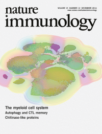
No. 12 December 2014
Myeloid cell subsets are difficult to classify objectively because of the range of subjective definitions based on various cell surface markers. Through the use of a 38-antibody mass cytometry analysis panel and a dimensionality-reduction method, Becher et al. (p 1181; and News and Views by Irish, p 1095) systematically categorize mouse myeloid cells from eight different tissues. The original figure by Jinmiao Chen shows individual myeloid cells from all mice and all tissues plotted on the basis of their phenotypic relationships in a single plot.Artwork by Lewis Long.
-
No. 11 November 2014
Cells of the immune system tailor their responses to microbe size. Branzk et al. demonstrate that neutrophils sense microbe size and selectively release neutrophil extracellular traps to control large pathogens (p 1017; News and Views by Mathew L. Wheeler and David M. Underhill, p 1000). The original image shows neutrophils responding to large fungal filaments (red) by releasing such traps that contain the antimicrobial protein myeloperoxidase (green) and decondensed DNA (blue). Image by Nora Branzk and Venizelos Papayannopoulos. Artwork by Lewis Long.
-
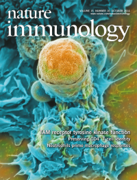
No. 10 October 2014
Billions of apoptotic cells are cleared from human tissues each day, during both tissue homeostasis and the resolution of inflammation. Lemke and colleagues show that the TAM receptor tyrosine kinases Axl and Mer are specialized to function as phagocytic receptors in these two different environments (p 920). The original image is a scanning electron micrograph of an inflammatory macrophage engulfing an apoptotic cell. Original image by Anna Zagórska and Matt Joens. Artwork by Lewis Long.
-
No. 9 September 2014
This month's Focus features a series of three specially commissioned Reviews and a Perspective that provide an in-depth analysis of signaling via the T cell antigen receptor and its regulation, as well as the functional consequences of the T cell antigen receptor's recognition of peptides presented on major histocompatibility complex molecules. See http://www.nature.com/ni/focus/TCRsignaling/index.htmlArtwork by Lewis Long depicts a TCR.
Focus
-
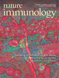
No. 8 August 2014
Cells of the immune system release cytokine 'factories' in the form of bioactive extracellular inflammasome assemblies. New findings by Franklin et al. and Baroja-Mazo et al. (pp 727 and 738; and News and Views by Broderick & Hoffman, p 698) identify extracellular functions of inflammasomes that are important for autoimmunity and the defense against pathogens. The original image shows the recruitment of neutrophils to injected extracellular specks of the adaptor ASC, which demonstrates their function as danger signals. Original image by Jacqueline M. Ratter. Artwork by Lewis Long.
-
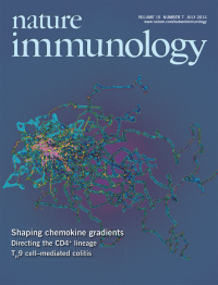
No. 7 July 2014
Proper trafficking of cells of the immune system and their positioning in lymphoid tissues requires chemotactic guidance. Rot and colleagues show that chemokine gradients are actively established and maintained in lymph node subcapsular sinus regions by the atypical chemokine receptor CCRL1 (p 623; and News and Views by Woodruff & Turley, p 595). Original image shows tracks of dendritic cell migration observed in vitro in response to gradients of the chemokine CCL19 shaped by CCRL1. Original image by Kathrin Werth. Artwork by Lewis Long.
-
No. 6 June 2014
Post-transcriptional and posttranslational modifications have profound influences on all aspects of immunity. Much like a blacksmith hammering a piece of metal into something functional, phosphorylation, methylation and acetylation can also alter NF-κB's function. This month's Focus features five specially commissioned Reviews that discuss the role of such modifications in various aspects of the immune system ranging from development to activation to immunopathology. www.nature.com/focus/ptm.Artwork by Lewis Long.
-
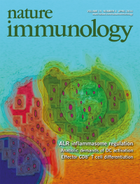
No. 5 May 2014
Treg cells suppress through a variety of mechanisms. Altman and colleagues show that CTLA-4-PKC-η interaction at the Treg cell immunological synapse is required for contact-dependent suppression by Treg cells (p 465; and News and Views by Wülfing, Tunbridge & Wraith, p 408). The original image shows GFP-tagged PKC-η (green) accumulated at the interface between a Treg cell (red) and an antigenpresenting B cell. Cyan indicates the nuclei. Original image by Tadashi Yokosuka and Takashi Saito. Artwork by Lewis Long.
-
No. 4 April 2014
AIM2 and IFI16 are intracellular, inflammasome-activating sensors of DNA that are pivotal for the detection of viral infections. Stehlik and colleagues (p 343; and News and Views by Eisenbarth, p 311) found a previously unrecognized protein, POP3, that functions as an inhibitor of DNA-induced inflammasomes. The original image, generated by Sonal Khare and Christian Stehlik, shows a macrophage transduced with a green fluorescent protein-expressing adenovirus (green) and DNA (blue) and stained for AIM2 (magenta). Artwork by Lewis Long.
-
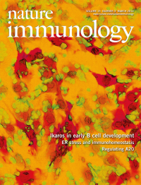
No. 3 March 2014
Loss of the transcription factor Ikaros is associated with a poor prognosis for certain leukemias. Georgopoulos and colleagues (p 294) show that loss of Ikaros arrests B lymphoid progenitors at an adherent and proliferative pre-B cell stage. The original image shows Ikaros-deficient pre-B cells (blue) with enhanced adhesion to bone marrow stroma (green) and more activity of the kinase FAK (red).Artwork by Lewis Long.
-
No. 2 February 2014
More detailed and comprehensive analysis of the immune system will reveal basic and translational insights and may lead to the development of new therapeutic strategies. This month's joint Focus features articles that discuss the new technologies and computational approaches that enable this sort of high-dimensional characterization of components of the immune system. www.nature.com/focus/high_dimensional_immune_analysisArtwork by Lewis Long.
-
No. 1 January 2014
The sensing of nucleic acids is pivotal for detecting viral infections. Reinecker and colleagues (p 63) show that the microtubule-associated protein GEF-H1 is activated after the intracellular detection of nucleic acids and is required for antiviral defense against RNA viruses. The original image, generated by Hans-Christian Reinecker and Hao-Sen Chiang, shows GEF-H1 (green) and the microtubule network (red); yellow indicates colocalization of GEF-H1 and a-tubulin.Artwork by Lewis Long.