Thank you for visiting nature.com. You are using a browser version with limited support for CSS. To obtain
the best experience, we recommend you use a more up to date browser (or turn off compatibility mode in
Internet Explorer). In the meantime, to ensure continued support, we are displaying the site without styles
and JavaScript.
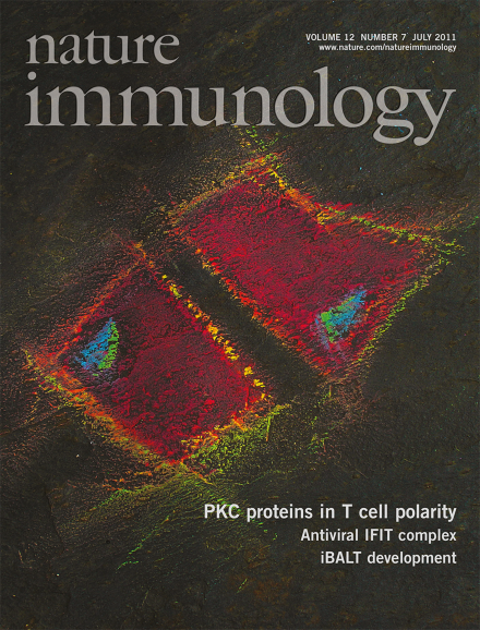
In T cells, three distinct protein kinase C (PKC) isozymes function sequentially to induce polarization of the microtubule-organizing center toward the immunological synapse, as reported by Huse and colleagues (p 647). Original image shows the PKC substrate Marcksl1 associated with the plasma membrane (red; total internal reflection fluorescence microscopy) and the microtubule-organizing center (blue; epifluorescence microscopy) in two T cells. Marcksl1 is depleted from the plasma membrane by PKC phosphorylation. Original image by Xin Liu. Artwork by Lewis Long.
This report describes advances in the understanding of how microbes elicit and evade immune responses and the sensing of pathogens by host cells that leads to the activation and production of intra- and extracellular signaling molecules.
For 50 years, immunologists have been meeting each winter in California to discuss new findings and theories in immunology. A recurring theme this year was the continuous conversation between the innate and adaptive arms of the immune system. To mark the 50th anniversary of this meeting, some of the speakers took a look back half a century to see how far immunologists have come.
Although the role of adaptive immunity in asthma is well characterized, there is relatively little understanding of the contribution of innate immunity to asthma. Studies now suggest that interleukin 13 produced by innate natural helper cells in the lungs has a substantial and underappreciated role in asthma exacerbation.
New data show that the interferon-induced gene product IFIT1 is a sensor for 5′-triphosphorylated RNA of viral origin and that it functions within a larger IFIT complex to inhibit viral replication.
In response to inhaled pathogens, lymphoid tissues can form in the lung. The driving force behind this organogenic process turns out to be interleukin 17–mediated activation of lung stromal cells.
The T cell antigen receptor is functionally coupled to many kinases and adaptor proteins. Analysis of the spatiotemporal organization of the T cell antigen receptor signaling cascade suggests that adaptor-containing intracellular vesicles are essential for proper signal propagation.
Tumor necrosis factor promotes potent inflammatory immune responses that can result in pathological tissue damage. Ivashkiv and colleagues show that it induces a regulatory feedback pathway that dampens inflammatory signaling via the kinase GSK3.
Type I natural killer T cells are characterized by an invariant Vα14-Jα18 T cell antigen receptor α-chain. Godfrey and colleagues describe a population of CD1d-restricted natural killer T cells that express a previously unidentified canonical Vα10-Jα50 α-chain.
The antiviral protein IFIT1 is upregulated substantially in the cytoplasm of virus-infected cells. Superti-Furga and colleagues show that IFIT1 is part of a virus-recognition pathway that sequesters specific viral RNA.
Innate lymphoid cells are present in the gut and support T helper type 2 responses. Umetsu and colleagues now identify these cells in the lung where they contribute to T cell– and B cell–independent asthma.
Ectopic bronchus-associated lymphoid tissue can form in lungs after pulmonary infection. Randall and colleagues show that aerosolized lipopolysaccharide induces the formation of this tissue in neonatal mice by a process dependent on interleukin 17.
Cognate recognition by T cells drives the formation of immune synapses. Huse and colleagues show that a sequential cascade of distinct protein kinase C isoforms is needed to reorient microtubule-organizing centers and to establish T cell polarity.
In quiescent T cells, the signaling adaptor Lat aggregates into cell-surface clusters. Gaus and colleagues demonstrate that pre-existing clusters of Lat do not participate in signaling from the T cell antigen receptor, which relies instead on recruitment of Lat from subsynaptic vesicles.
The transcriptional control of T cell exhaustion remains unclear. Wherry and colleagues show that the transcription factor T-bet regulates CD8+ T cell exhaustion and inhibitory receptor expression.
The environmental cues involved in regulating germinal center size are not fully understood. Cyster and colleagues show that the sphingosine 1-phosphate receptor S1P2 controls the survival and localization of B cells in germinal centers by antagonizing signaling by the kinase Akt and follicular chemoattractants.