Thank you for visiting nature.com. You are using a browser version with limited support for CSS. To obtain
the best experience, we recommend you use a more up to date browser (or turn off compatibility mode in
Internet Explorer). In the meantime, to ensure continued support, we are displaying the site without styles
and JavaScript.
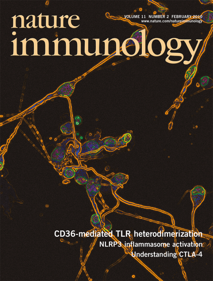
Neurons in in vitro culture are exposed to inflammatory mediators released by β-amyloid-activated microglial cells. Moore and colleagues (p 155) show that a CD36-TLR4-TLR6 heterotrimer mediates β-amyloid recognition and microglial cell activation in the brain. The original confocal image shows CAD mouse neuronal cells cultured together with microglial cells stimulated with Aβ1–42 and stained with antibody to neuronal class III β-tubulin (green). DAPI nuclear staining is blue. Original image by Janine M van Gils and Laurent Boyer. Artwork by Lewis Long.
The signaling intermediates that activate inflammasomes are elusive. New data now show that reactive oxygen species causes TXNIP to associate with NLRP3 and activate the inflammasome.
The mutator activation-induced cytidine deaminase is essential for immunoglobulin diversification but can be detrimental in other settings. A new comprehensive analysis investigates how its gene expression is regulated.
CTLA-4 is a potent coinhibitory molecule that is critical for peripheral T cell tolerance. New data suggest that CTLA-4 exerts its critical immunoregulatory functions by controlling antigen-specific conventional T cells as well as regulatory T cells.
Preclinical studies in mice suggest that the natural killer cell receptor NKp46 may be a new molecular target for delaying the progression of type 1 diabetes by interfering with unexpected natural killer cell–mediated recognition of pancreatic beta cells.
Natural killer cells infiltrate the pancreas during type 1 diabetes. Mandelboim and co-workers find that the natural killer receptor NKp46 recognizes ligands on pancreatic beta cells and is essential for full diabetes development.
CTLA-4-deficient mice develop a lethal multiorgan lymphoproliferative disorder. Murphy and colleagues definitively demonstrate that at least some of the pathogenic T cells that drive this disorder show reactivity to nonlymphoid tissue self antigens.
The signaling intermediates that activate inflammasomes have remained elusive. Tschopp and co-workers now describe that a thioredoxin-binding protein is a reactive oxygen species–regulated component of the NLRP3 inflammasome.
Toll-like receptor (TLR) signaling activates the transcription factor NF-κB and production of proinflammatory cytokines. O'Neill and colleagues show that TLR signaling induces the microRNA miR-21 to dampen PDCD4 expression, which leads to less NF-κB activity and more IL-10 production.
Immunoglobulin diversification is absolutely dependent on the action of activation-induced cytidine deaminase, which must be tightly controlled. Honjo and colleagues systematically analyze the regulatory elements that govern expression of the gene encoding this deaminase.
The molecular mechanisms behind recognition of altered self remain unclear. Moore and co-workers show that oxidized low-density lipoprotein (LDL) and β-amyloid trigger inflammatory signaling through a heterodimer of Toll-like receptors 4 and 6.
Pre-TCR signaling is essential for the passage of thymocytes through the β-selection checkpoint. Ravichandran and co-workers find that CXCR4 acts as a costimulator for the pre-TCR and is needed for optimal progression through β-selection.
B cell development requires interleukin 7 signals that activate the transcription factor STAT5. Busslinger and colleagues report that STAT5 has a permissive rather than instructive role in pro-B cell survival and immunoglobulin recombination.