Thank you for visiting nature.com. You are using a browser version with limited support for CSS. To obtain
the best experience, we recommend you use a more up to date browser (or turn off compatibility mode in
Internet Explorer). In the meantime, to ensure continued support, we are displaying the site without styles
and JavaScript.
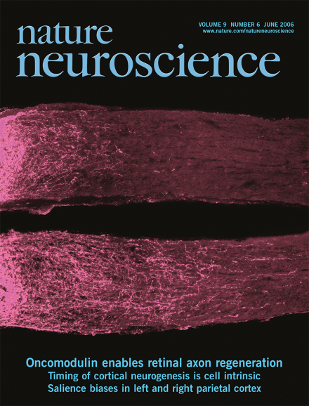
The optic nerve does not regenerate after injury in mature mammals, but retinal ganglion cells do regenerate lengthy axons beyond the site of optic nerve injury when stimulated by macrophage activation in the eye. Benowitz and colleagues now show that the Ca2+ binding protein oncomodulin is a macrophage–derived growth factor for retinal ganglion cells and other neurons. The cover image shows two injured optic nerves, the bottom one from an animal that has been treated with oncomodulin plus a cAMP stimulant, resulting in robust axon regeneration. (pp 715 and 843)
Macrophages near neuronal cell bodies can promote regeneration in an otherwise inhibitory environment. Now Yin et al. identify oncomodulin as a factor secreted from macrophages that promotes extensive regeneration of lesioned optic nerve axons when applied together with elevated cyclic AMP (cAMP) and mannose.
During development, neurons destined for different neocortical layers are sequentially generated. Shen et al. report that this timing is programmed within individual progenitor cells and depends mainly on cell-intrinsic mechanisms.
Dendritic spines confine activity-dependent changes to the affected synapses. A paper in this issue reports that retarded diffusion in dendrites of aspiny interneurons may create dendritic biochemical compartments without spines.
Exactly how animals switch between different sleep states remains unknown. A new study in Nature provides a glimpse into the mechanisms and anatomy of the brain regions that trigger rapid eye movement sleep.