Volume 26
-
No. 12 December 2023
Mouse model amyloid structuresMice that have been genetically modified to mimic Alzheimer's disease are vital for research into the condition. Zielinski et al. analyzed amyloid-β fibrils in different mouse models and identified striking structural variations. The image shows a cross-section through a 3D reconstruction of an amyloid-β fibril from an APP23 mouse, identical to the type 2 fibril found predominantly in patients with familial Alzheimer's disease (a rare, early-onset inherited form).
See Zielinski, Peralta Reyes et al.
-
No. 11 November 2023
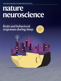
Brain and behavioral responses during sleepSleep is assumed to be a state of behavioral disconnection with the outside world, but Türker and colleagues report that human sleepers can intermittently frown or smile in response to verbal stimuli across most sleep stages. This observation challenges the current definition of sleep. The cover image symbolically depicts the idea that a sleeping person can still process information from the external environment.
See Türker, Munoz Musat, Chabani et al.
-
No. 10 October 2023
Sensorimotor striatum and complex motor sequencesThe cover art illustrates the ubiquitous process of producing complex motor sequences, exemplified by the pianist. How such feats are implemented in neural circuitry is addressed by Mizes et al. in this issue. They study rats performing a ‘piano task’, contrasting the role of the basal ganglia in overtrained sequences with ones assembled in response to visual cues, like notes on a musical staff. They find that the basal ganglia help to shape low-level movement kinematics for both types of sequence but are dispensable for high-level sequencing of cue-guided behaviors.
See Mizes et al.
-
No. 9 September 2023
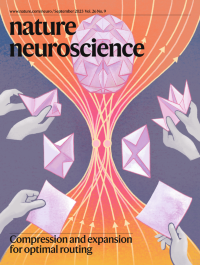
Compression and expansion for optimal routingIn origami, flat paper sheets are first folded and then unfolded to form beautiful three-dimensional figures. The authors describe an analogous principle by which sensorimotor signals are compressed and then expanded to form high-dimensional representations in the cerebellum and similar structures.
See Muscinelli et al.
-
No. 8 August 2023
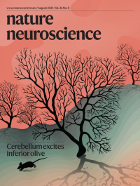
Cerebellum excites inferior oliveWang and colleagues have identified an excitatory cerebello-olivary pathway and its role in fine motor control. The cover art depicts a mouse running vigorously through a landscape of Purkinje cell trees on a rugged mountain, showcasing the intricate gyri and sulci of the cerebellum. A sunrise scene bathes the entire composition, casting light on the Purkinje cell trees and forming shadows resembling olive trees. This artwork symbolizes the profound importance of cerebello-olivary excitation in controlling movements, which lies at the core of this study.
See Wang et al.
-
No. 7 July 2023
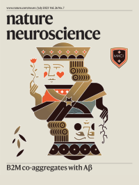
B2M co-aggregates with AβIn this issue, Zhao et al. demonstrate a noncanonical role of B2M in the pathogenesis of Alzheimer’s disease. B2M is a key component of major histocompatibility complex class I (MHCI), which is crucial for host defense against pathogen infection. In addition to forming a complex, B2M can dissociate from MHCI. Secreted B2M can cross the blood–brain barrier and co-aggregate with β-amyloid in the brain. The image illustrates the two different sides of B2M–MHCI in the immune and nervous systems. In the immune system, B2M–MHCI (king at the top) prevents pathogen infection and safeguards our health (shield). However, in the brains of people with Alzheimer’s disease, secreted B2M (king at the bottom) can impair neuronal function.
See Zhao et al.
-
No. 6 June 2023
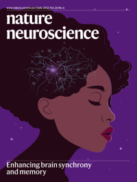
Enhancing brain synchrony and memoryThe cover art depicts brain activity during human sleep. It is based on an article from Geva-Sagiv et al., which describes how intracranial stimulation precisely timed with slow-wave activity in the medial temporal lobe during sleep enhances coupling of neuronal oscillations across regions and improves memory performance.
See Geva-Sagiv et al.
-
No. 5 May 2023
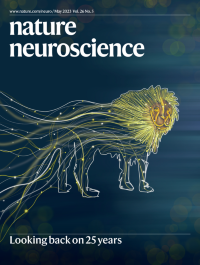
Looking back on 25 yearsThe cover image is a depiction of connections of past neuroscience research coming together to create forward momentum in the form of a lion. The lion represents a Sanskrit phrase for retrospection — सिंहावलोकन or sinhavalokan — which translates to “as a lion looks back.” It originates from the behavior of a lion pausing momentarily to look back at the path traversed and forward to what lies ahead. As Nature Neuroscience completes 25 years, we pause and reflect on how we have worked with the neuroscience community in communicating the most impactful research. Cover concept: Marina Corral Spence and Sachin Ranade.
See Editorial
-
No. 4 April 2023
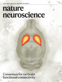
Consensus for rat brain functional connectivityTwo hundred investigators across nearly fifty centers developed StandardRat, a consensus functional MRI acquisition protocol for estimating functional connectivity in the rat brain.
See Grandjean et al.
-
No. 3 March 2023
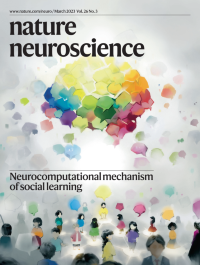
Neurocomputational mechanism of social learningJiang et al. identify a neurocomputational mechanism by which the human brain biases the integration of information transmitted on social networks. The cover art illustrates that the brain aggregates diverse information received from other individuals embedded on a social network.
See Jiang et al.
-
No. 2 February 2023
Self and other coding in batsOmer et al. report two distinct populations of hippocampal time cells in bats — one encoding time × context and another encoding pure time — as well as time cells that encode time and context for another animal; these neurons might underlie the perception of interval timing and episodic memory for self and other.
See Omer et al.
-
No. 1 January 2023
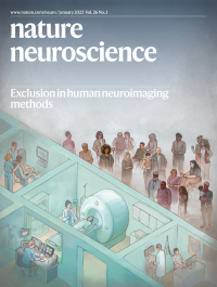
Exclusion in human neuroimaging methodsThe use of field-standard approaches in neuroscience and psychology can exclude participants from research, biasing our understanding of brain–behavior relations. Ricard, Parker, and colleagues discuss how we might address inequity in our scientific methodology. The cover image is a stylized illustration depicting exclusion in human neuroimaging methods. Cover concept: Mona Li, Jocelyn Ricard. Printed with permission from Mona Li Visuals.
See Ricard, Parker et al.