Thank you for visiting nature.com. You are using a browser version with limited support for CSS. To obtain
the best experience, we recommend you use a more up to date browser (or turn off compatibility mode in
Internet Explorer). In the meantime, to ensure continued support, we are displaying the site without styles
and JavaScript.
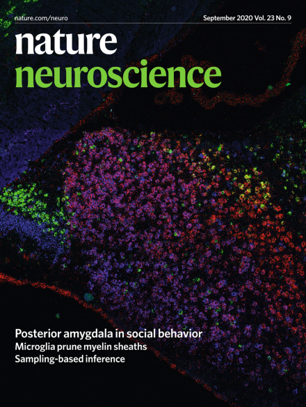
Yamaguchi et al. identify two largely spatially distinct subpopulations in the posterior amygdala (PA): MPN-projecting PA cells in the dorsomedial part and VMHvl-projecting PA cells in the ventrolateral part. The cover shows RNA expression patterns of genes enriched in whole PA (red: Zic2), PAMPN (blue: Npy2r) and PAVMHvl (green: Calb2) cells.
Pathological tau disrupts the association between nitric oxide (NO) synthase and PSD95, impairing NO signaling and neurovascular coupling before causing neurodegeneration. Stopping production of pathological tau rescues NO signaling, neurovascular coupling and neuronal function, but doesn’t remove tangles, suggesting that (like amyloid-β) soluble tau is an important driver of early neurovascular dysfunction and subsequent neuronal damage.
Sleep is controlled by a cocktail of neurotransmitters, but it is difficult to measure these in the brain. A new study by Tamaki et al. reveals how the balance between excitation and inhibition oscillates as the brain moves through sleep stages and how this impacts upon memory consolidation and stabilization.
Microglia refine the developing CNS by engulfing excess neurons and synapses. Hughes and Appel here show that microglia also prune myelin sheaths in a neuronal activity-regulated manner to sculpt developmental myelination.
The complement–microglia pathway is a key mediator of synapse elimination in development and disease. Cong et al. show that neurons endogenously express a complement inhibitor, SRPX2, that regulates synapse elimination in development.
Park et al. demonstrate in tauopathy models that tau disrupts the interaction between neuronal nitric oxide synthase and PSD95, uncoupling glutamatergic synaptic activity from nitric oxide production and dampening the hemodynamic response to activation.
Most studies of autism spectrum disorder (ASD) have focused on neuronal mechanisms. Here, the authors describe vascular impairments in a mouse model of 16p11.2 deletion syndrome using physiological and genetic approaches to examine endothelial-dependent phenotypes.
Kelly et al. describe two cerebellum–thalamus–mPFC pathways in mice that regulate social and repetitive behavior. PC activation in Rcrus1 and posterior vermis improved social and reduced repetitive behaviors, respectively, in PC-Tsc1 mutant mice.
Yamaguchi et al. identify a little-known amygdalar region, the posterior amygdala, as a key node in male mouse social behaviors. Two largely non-overlapping subpopulations in the posterior amygdala form parallel projections to distinct hypothalamic regions to regulate mating and fighting.
Charlet, Grinevich et al. show that social touch between female rats activates parvocellular oxytocin neurons; these neurons control social behavior by coordinating the responses of the much larger population of magnocellular oxytocin neurons.
Neural oscillations, transients and variability are widely observed in sensory cortices. All these features emerge in neural networks optimized for the singular task of representing perceptual uncertainty in the variability of neural responses.
Tamaki et al. measured MRS changes in sleeping humans trained on a visual task. During NREM sleep, learning gains were associated with enhanced visual cortical plasticity that was also seen independent of learning. REM sleep stabilized plasticity only after pre-sleep learning.
Deschloroclozapine (DCZ) is a broadly useful chemogenetic agonist for studies using nonhuman primates and mice. DCZ rapidly and reversibly activates DREADDs, and its binding can be visualized noninvasively by positron emission tomography.
SPARC is an all-genetic toolkit to express effectors in precise proportions of neurons. This method enables imaging of individual neurons and manipulation of neuronal subpopulations.