Volume 34
-
No. 12 December 2016
High-resolution, live imaging of an entire developing Drosophila embryo at different stages, with adaptive light-sheet microscopy, highlighting the nuclei of all of the embryo's cells in blue and its developing nervous system in red and yellow. Royer et al. present an approach to continuously evaluate changes in the embryo's optical properties and to adapt the microscope's configuration to these dynamic conditions to recover and maintain high spatial resolution (p 1267). Image credit: Julia Kuhl, Loïc A. Royer & Philipp J. Keller
-
No. 11 November 2016
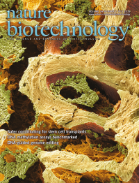
Colored scanning electron micrograph of a colon cancer stem cell. Image credit: Martin Oeggerli / Micronaut, supported by School of Life Sciences, FHNW
-
No. 10 October 2016
A microfuidics array of connected suspended microchannel resonators separated by serpentine channels to measure cellular growth rate. The channels allow the cells time to grow as they flow through the cantilevers, before they are weighed with high precision. Cermak et al. use the approach to examine growth rate in bacteria, yeast and mammalian cells at the single-cell level (p 1052). Image credit: Selim Olcum & Nathan Cermak
-
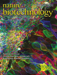
No. 9 September 2016
Mouse hippocampal neurons expressing membrane-anchored fluorescent proteins. Tillberg et al. (p 987) and Ku et al. (p 973) improve expansion microscopy approaches to enable super-resolution image quality with off-the-shelf reagents. Image credit: Fei Chen, Dawen Cai & Edward Boyden
-
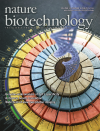
No. 8 August 2016
Artist's impression of an experiment establishing the specificity patterns of the Cpf1 RNA-guided nuclease. Kim et al. (p 875) and Kleinstiver et al. (p 881) analyze the genome-wide on-target and off-target specificities of Cpf1 in vitro and in human cells. Image credit: IBS, Institute for Basic Science, Seoul, South Korea
-
No. 7 July 2016
A scanning electron micrograph of bone tissue. The bone marrow (orange) provides the niche for hematopoietic stem cells. Palchaudhuri et al. describe an antibody-based agent for depleting cells in the bone marrow before hematopoietic stem cell transplantation (p 738). Image credit: Steve Gschmeissner, Science Source
-
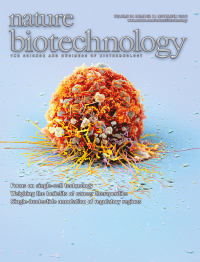
No. 6 June 2016
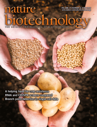
Crop losses caused by plant pathogens are managed by either planting resistant varieties or using pesticides. A trio of papers from The Sainsbury Laboratory in Norwich, UK, provides evidence that engineering pathogen resistance could provide sustainable genetic solutions to crop pests and pathogens. In one report, soybean is made resistant to Asian soybean rust using a gene cloned from the orphan legume pigeonpea (p 661). In two accompanying reports, resistance genes for potato late-blight and wheat stem rust are identified by developing rapid resistance-gene cloning methods (pp 656 and 652). Image credit: Andrew Davis, John Innes Centre, Norwich, UK; The hands are from: Kamik Witek (potato), Burkhard Steuernagel (wheat) and Kim Wood (soybean).
-
No. 5 May 2016
Artist's representation of individuals (highlighted in green) who remain healthy despite carrying mutations that cause severe Mendelian diseases. Chen et al. analyze genetic data from over half a million people and identify 13 healthy individuals who harbor highly penetrant disease-causing mutations. No study participants are depicted (p 531). Image credit: Marina Spence; images provided by John Ambrose
-
No. 4 April 2016
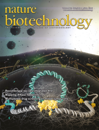
Artist's impression of a recombinase removing an HIV provirus from the genome of a T cell. Karpinski et al. have engineered a recombinase that excises integrated HIV genomes from the genome of host cells with very high specificity and is effective against the majority of clinical isolates (p 401). Credit: Heinrich Pette Institute
-
No. 3 March 2016
Nature Biotechnology celebrates its 20-year anniversary. Cover art: Erin Boyle
-
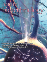
No. 2 February 2016
Artist's impression of a patch clamp pipette probing the interior of a single neuron in brain tissue. In this issue Cadwell et al. (p 199) and Fuzik et al. (p 175) present methods for the combined electrophysiological, transcriptomic and morphologic profiling of single cells in situ. Credit: Sputnik Animation, Ed Boyden and the McGovern Institute for Brain Research at MIT
-
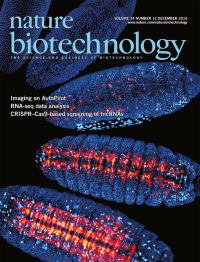
No. 1 January 2016
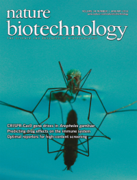
A newly emerged female Anopheles gambiae mosquito is shown, resting on water before its first flight. Andrew Hammond and colleagues (p 78) develop a CRISPR-Cas9-based gene drive system that targets female fertility in Anopheles gambiae, the main vector for the malaria parasite. This paves the way for the development of efficient gene drives that could suppress mosquito populations. Photographers: Andrew Hammond and Alekos Simoni.