Volume 22
-
No. 12 December 2004
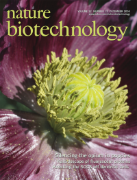
The flower of the opium poppy, Papaver somniferum, showing the sexual parts. Allen et al. have used RNA interference to modify the alkaloid content of this poppy (see p. 1559). Credit: Jeremy Burgess/Photo Researchers, Inc.
-
No. 11 November 2004
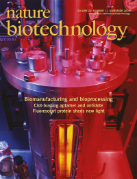
Pilot fermentation vat for the culture of microorganisms. As well as advances in the engineering of cells for protein production, new and more efficient fermentation systems are being designed by engineers. Credit: E. Young/Photo Researchers Inc.
-
No. 10 October 2004
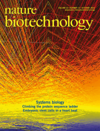
The cover shows computer analysis of DNA array data comparing the expression patterns of 22500 human genes in normal and cancerous prostate cells. Red represents high levels of gene expression and blue represents low levels. Picture by Veronique Blanc and Qin Wang from Wellcome Trust Medical Photographic Library.
Focus
-
No. 9 September 2004
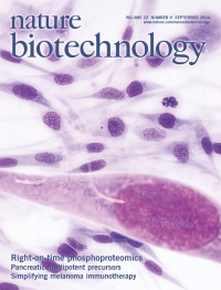
Representation of the epidermal growth factor receptor (EGFR) pathway HeLa cells. The dynamics of EGFR signaling have been studied using a mass spectrometry-based proteomics method capable of analyzing the phosphoproteome at multiple time points (see Blagoev et al., p. 1139). Photo courtesy of Richard J. Green/Photo Researchers, Inc.
-
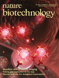
No. 8 August 2004
Representation of quantum dots, optimized for in vivo delivery, traveling through a blood vessel to bind specifically to receptors on tumor cells (foreground). Such particles promise to significantly advance cancer imaging (see Nie and colleagues, p. 969). Graphic by Ken Eward © Biografx.
-
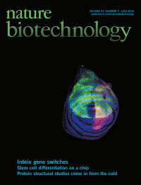
No. 7 July 2004
Drosophila wing imaginal disc showing repression of Gal4 expression by a temperature-sensitive intein-Gal80 construct. The posterior compartment is labeled with anti-Engrailed (red), DNA is stained with DAPI (blue) and expression of the Gal4-GFP construct (green) is shown to be repressed on the anterior side of the compartment. On page 871, Perrimon and colleagues report on the post-transcriptional regulation of protein synthesis in Drosophila using temperature-sensitive inteins.
-
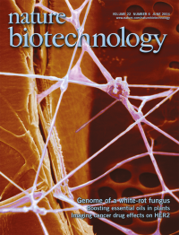
No. 6 June 2004
1000X SEM image of a longitudinal section of Phanerochaete chrysosporium colonizing aspen. Hyphae are visible throughout and in the vessel pit on the right. Photo courtesy of T. Kuster (USDA Forest Products Laboratory, Madison, WI). On page 695, Cullen and colleagues report the full genome sequence of the white-rot fungus P. chrysosporium.
-
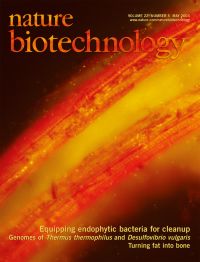
No. 5 May 2004
A root of a poplar colonized by the green fluorescent protein-labeled endophytic bacterium Pseudomonas putida VM1453. Photo courtesy of Kieran Germaine, David Ryan and David Dowling Department of Applied Biology & Chemistry, Institute of Technology, Carlow, Ireland). On page 583, van der Lelie and colleagues describe a yellow lupin containing a modified endophytic symbiont capable of toluene degradation.
-
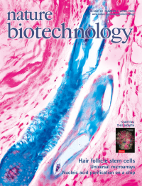
No. 4 April 2004
Isolated hair follicle stem cells form all components of the cutaneous epithelium, including new hair follicles, hairs, epidermis and sebaceous glands, when injected together with mesenchymal cells into immunodeficient mice. The hair follicle stem cells were isolated from Rosa reporter mice expressing lacZ in every cell (Morris et al., p 411).
-
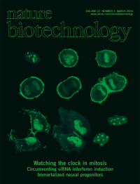
No. 3 March 2004
Images of Hela cells expressing a live cell fluorescent mitosis biosensor as they go through cell division. The images start with a representative cell in G2, (upper left panel), and show examples of the localization of the biosensor in cells as they proceed through the mitotic stages, prometaphase, metaphase, anaphase, telophase, and cytokinesis. (See Meyer and colleagues, p 306). Image courtesy of Joshua Jones.
-
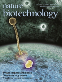
No. 2 February 2004
Representation of a bacteriophage docking on a target molecule in a bacterial membrane. The harnessing of bacteriophage antimicrobial strategies to identify novel bactericidal mechanisms allows the design of assays to screen small molecule libraries for compounds mimicking those effects (see Liu et al., p 185). Electron microscopy images of Staphylococcus aureus phage 77, courtesy of Dr. Hans-Wolfgang Ackermann, Department of Medical Biology, Laval University, Quebec, Canada. Artwork by InViVo Communications Inc. Toronto, Canada.
-
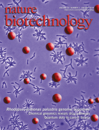
No. 1 January 2004
Characteristic, reddish colonies of the purple photosynthetic bacterium Rhodopseudomonas palustris superimposed on a picture of its rod-shaped cells visualized under the light microscope. This issue reports the complete sequence of R. pulustris, a metabolically highly versatile microorganism (see pp 55-61).