Volume 508
-
No. 7497 24 April 2014
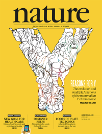
Mammalian Y chromosomes, known for their roles in sex determination and male fertility, often contain repetitive sequences that make them harder to assemble than the rest of the genome. To counter this problem Henrik Kaessmann and colleagues have developed a new transcript assembly approach based on male-specific RNA/genomic sequencing data to explore Y evolution across 15 species representing all major mammalian lineages. They find evidence for two independent sex chromosome originations in mammals and one in birds. Their analysis of the Y/W gene repertoires suggests that although some genes evolved novel functions in sex determination/spermatogenesis as a result of temporal/spatial expression changes, most Y genes probably persisted, at least initially, as a result of dosage constraints. In a parallel study, Daniel Bellott and colleagues reconstructed the evolution of the Y chromosome, using a comprehensive comparative analysis of the genomic sequence of XY gene pairs from seven placental mammals and one marsupial. They conclude that evolution streamlined the gene content of the human Y chromosome through selection to maintain the ancestral dosage of homologous XY gene pairs that regulate gene expression throughout the body. They propose that these genes make the Y chromosome essential for male viability and contribute to differences between the sexes in health and disease. Cover: Daren Newman
-
No. 7496 17 April 2014
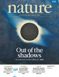
Solar Eclipse, 1932, by Howard Russell Butler (1856�1934). The first of the two solar eclipses of 2014 occurs on 29 April. Unusually, much of the Moons shadow will miss Earth entirely, but the partial phases will be visible from Australia and parts of Antarctica. Plenty of photographs will be taken, tweeted and instagrammed, but will they be as evocative as the artists� efforts in the pre-photographic era? In an essay in this issue, Jay Pasachoff and Roberta Olson survey solar art from the Renaissance to the twentieth century and celebrate Howard Russell Butler as a star in the eclipse-painting firmament. Credit: Princeton University Art Museum/Art Resource NY/Scala, Florence
-
No. 7495 10 April 2014
Axonal projection patterns from 21 distinct cortical areas (differentially colour coded) derived from 21 mapping experiments to sample the entire cortex, and rendered in 3D by the Brain Explorer program. In this issue, Hongkui Zeng and colleagues present the first brain-wide, mesoscale connectome for a mammalian species � the laboratory mouse � based on cell-type-specific tracing of axonal projections. The wiring diagram of a complete nervous system has long been available for a small roundworm, but neuronal connectivity data for larger animals has been patchy until now. The new 3D Allen Mouse Brain Connectivity Atlas is a whole-brain connectivity matrix that will provide insights into how brain regions communicate. Much of the data generated in this project will be of relevance to investigations of neural networks in humans.
-
No. 7494 3 April 2014
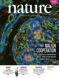
Basal mammary tumour cells from a donor mouse (red) expressing a lineage marker (green) comingle with host-derived epithelial cells. The elongated mammary duct on the left retains a normal bilayered architecture in the absence of infiltration by the donor-derived tumour subclone. Tumours often display a complex subclonal organization. In a mouse model of breast cancer initiated by Wnt signalling, Allison Cleary et al. show that some tumours are biclonal � composed of basal and luminal clones with distinct genetic alterations. These clones cooperate to maintain tumour growth that is dependent on secretion of Wnt by the luminal cells. When Wnt production is blocked, basal cells carrying Hras mutations recruit other Wnt-producing cells to restore tumour growth, or one of the original clones may acquire alternative means of activating the pathway. These findings illuminate how complex cellular interactions in heterogeneous tumours might sway treatment outcomes. On the cover, Cover: Thomas Abrahams/Penn State Imaging Core.