Thank you for visiting nature.com. You are using a browser version with limited support for CSS. To obtain
the best experience, we recommend you use a more up to date browser (or turn off compatibility mode in
Internet Explorer). In the meantime, to ensure continued support, we are displaying the site without styles
and JavaScript.
This issue highlights a nanopatterned microfluidic chip for the detection of circulating exosomes in patient samples, a microfluidic assay for the quantification of the metastatic propensity of breast cancer cells, an amplification-free electrical biosensor for nucleic acids, virtual staining of unlabelled tissue sections via deep learning, and a quantitative microimmunohistochemistry assay for the grading of immunostains on tumour tissues.
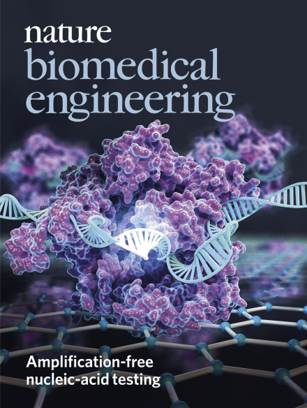
The cover illustrates an electrical biosensor for nucleic acids that relies on the binding of target sequences to Cas9 immobilized on a graphene field-effect transistor.
An electrical biosensor that relies on the binding of target nucleic acid sequences to Cas9 immobilized on a graphene field-effect transistor enables the rapid detection of mutations in purified samples without the need for nucleic acid amplification.
Early-stage ovarian cancer can be detected in few-microlitre plasma samples via a microfluidic chip, patterned with nanoporous herringbone structures so as to enhance the capture of extracellular vesicles from the samples.
A microfluidic assay that quantifies the abundance and degree of proliferation of migratory cells predicts the metastatic potential of breast-cancer cell lines and patient-derived cells.
An electrical biosensor combining CRISPR–Cas9 and a graphene field-effect transistor detects target genes in purified genomic samples at high sensitivity, within 15 minutes, and without the need for amplification.
A microfluidic chip with self-assembled 3D herringbone nanopatterns detects, with high sensitivity and specificity, tumour-associated exosomes in few-microlitre plasma samples from patients.
A microfluidic assay predicts the metastatic potential of breast cancer specimens by quantifying the abundance and proliferative index of the migratory cells within them.
Deep learning can be used to virtually stain autofluorescence images of unlabelled tissue sections, generating images that are equivalent to the histologically stained versions.
A quantitative microimmunohistochemistry assay based on the evolution of immunohistochemistry signals during tissue staining enhances the stratification of tumour samples.