Volume 26
-
No. 12 December 2021
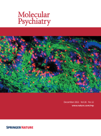
Cover image for Molecular Psychiatry, involving neurons being generated within patient-derived 3D dorsal-forebain organoids. For more information see the article by Notaras et al. on pages 7760-7783.
-
No. 11 November 2021
Immunofluorescent detection of TH+ (green) neurons and phospho-eIF2α (p-eIF2α; red) expression in SNc and VTA DA neurons of eIF2α(S51A/S51A) DAT-Cre and WT DAT-Cre mice, confirming positive targeting of dopaminergic (TH+) neurons for the expression of phospho-mutant eIF2α (scale bars represent 50 µm). Arrows indicate dopaminergic neurons (green) and p-eIF2α (red) co-stain. For more information see the article by Longo et al. on pages 6427-6450.
-
No. 10 October 2021
Blast-associated tauopathy in humans and rats with neurobehavioral syndromes. Left panel shows a positron emission tomography (PET) scan of the brain of a military veteran with a history of multiple blast exposures imaged with the PET tau ligand [18F]AV1451 (flortaucipir). Note the cortical ligand retention (red) at the grey/white matter junction, a distribution characteristic of the abnormal tau deposition found in chronic traumatic encephalopathy (CTE). Right panel shows perivascular deposition of hyperphosphorylated tau (green) in an astroglial process from a rat exposed to repetitive low-level blast exposures. Perivascular tau deposition is also a pathological feature of human CTE. For more information see the article by Dickstein et al. on pages 5940-5954.
-
No. 9 September 2021
Left-hand upper panel (low magnification) and left-hand lower panel (higher magnification): periventricular endothelial cells showed robust CD31 expression upon addition of WNT7a and GABA from day 0-5 of differentiation, while addition of GABA alone, selectively increased CD31 expression on endothelial cell-cell junctions. Center upper panel (low magnification) and center lower panel (higher magnification): co-labeling with endothelial cell markers, vWF (red) and CD31 (green) of human periventricular endothelial cells. Right-hand panel: high magnification image of vWF labeling of periventricular endothelial cells. For more information see the article by Debkanya Datta et al. on pages 4864–4883.
-
No. 8 August 2021
WT1 regulates the differentiation of human neural progenitor cells (hNPCs). Human NPCs generated from embryonic stem cells were infected with control or WT1-shRNA lentivirus and then neuronal differentiation was induced. Lentivirus infected hNPCs (green) were labeled with neuronal marker, TUJ1 (red). The number of TUJ1+ neurons is decreased in WT1-shRNA infected hNPCs (bottom panel). For more information see the article by Fen Ji et al. on page 4221–4233.
-
No. 7 July 2021
Immunofl uorescent staining of rat primary neurons (DIV. 14). Images show neuronal TrkB and APP co-localized on the cell surface. Neurons are labeled green with APP, red with TrkB, and blue with DAPI. For more information see the article by Yiyuan Xia et al. on page 2943–2963.
-
No. 6 June 2021
Immunohistochemical localization of TSPO protein in neuronal and non-neuronal hippocampal cells of adult mice using immunofluorescence staining. The photomicrographs show representative Z-stack images acquired through confocal microscopy, with nuclear staining (DAPI) in blue, TSPO in green and various CNS cells of interest in red. TSPO co-localizing with the cellular markers of interest appears in yellow. Top row images show TSPO co-localizing with the post-mitotic neuronal markers NeuN (left image), SMI-32 (middle image) and MAP-2 (right image). Lower row images show non-neuronal TSPO protein expression, including expression in Iba1-positive microglia (left image), GFAP-positive astrocytes (middle image) and Glut1-positive vascular endothelial cells (right image).For more information see the article by Tina Notter et al. on page 2025-2037.
-
No. 5 May 2021
Immunofluorescent staining of neuron-microglia co-cultures. Images show neurons from Pten WT/WT and from Pten autism mouse model Pten WT/m3m4 , and Pten m3m4/m3m (Left to right) co-cultured with Pten WT/WT microglia (Top panels) and Pten m3m4/m3m microglia (Bottom panels). Neurons are labeled green with Tuj1; C1q (red) punctae show accumulation on neuronal surfaces. For more information see the article by Prof. Charis Eng et al. on page 1458-1471.
-
No. 4 April 2021
Expression of the immediate early gene Arc (red) in mature (green) and newborn (green and blue) dentate granule neurons in mice with chronic DSS colitis during acquisition of spatial learning. For more information see the article by Katia P Karalis et al. on page 1248-1263.
-
No. 3 March 2021
Immunostaining of the neuronal marker beta III tubulin (green) and the transcription factor nuclear factor kappa B p65 (NF-κB, also known as RelA, red) in the LUHMES human neuronal cell line at differentiation day 2 (upper panels) and day 6 (lower panels). Nuclei are stained with Hoechst (blue). Inactive NF-κB is held in the cytoplasm by its inhibitor, IκB (left panels). Upon activation, IκB is phosphorylated by IκB kinase (IKK), and NF-κB is able to translocate into the nucleus, resulting in a pink color in LUHMES cells exposed to a mixture of inflammatory cytokines (middle panels). Pretreatment with the IKK inhibitor TPCA-1 largely prevents NF-κB nuclear translocation following cytokine exposure (right panels). For more information see the article by Prof. Pamela Lein et al. on pages 875-887.
-
No. 2 February 2021
Cell-specific increased surface P2X4mCherryIN in P2X4 internalization-defective knock-in mice. Surface P2X4 (green) is almost absent in MAP2-positive hippocampal neurons (magenta) of control mice expressing wild-type P2X4 but highly increased at the surface of CaMK2Cre-P2X4mCherryIN mice expressing non-internalized P2X4mCherryIN in excitatory neurons (first, second column). Middle, enlarged image of a neuronal dendrite, showing intracellular (red) and surface clustering of P2X4mCherryIN (green) at extra- and post-synaptic regions indicated by Homer 1c (blue). Right column, P2X4mCherryIN (red) is expressed in GFAP-positive (green) astrocytes from hippocampal cultures of CMVCre-P2X4mCherryIN mice. For more information see the article by Eléonore Bertin et al. on pages 629-644.
-
No. 1 January 2021
Single molecule fluorescence in situ hybridization showing Mineralocorticoid Receptor (Nr3c2) expression in mouse hippocampal area CA2. Left: Nr3c2 expression (magenta) in CA1 (top inset) compared with CA2 (bottom inset). Right: Same as left but also including nuclei (blue) and CA2 markers Pcp4 (green) and Acan (red). For more information see the article by Dr. Serena Dudek et al. on pages 350-364.