
Volume 26 Issue 9, September 2021
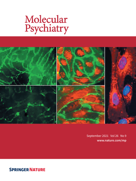
Left-hand upper panel (low magnification) and left-hand lower panel (higher magnification): periventricular endothelial cells showed robust CD31 expression upon addition of WNT7a and GABA from day 0-5 of differentiation, while addition of GABA alone, selectively increased CD31 expression on endothelial cell-cell junctions. Center upper panel (low magnification) and center lower panel (higher magnification): co-labeling with endothelial cell markers, vWF (red) and CD31 (green) of human periventricular endothelial cells. Right-hand panel: high magnification image of vWF labeling of periventricular endothelial cells. For more information see the article by Debkanya Datta et al. on pages 4864–4883.
Image
-
Advertisement
