
Volume 26 Issue 2, February 2021
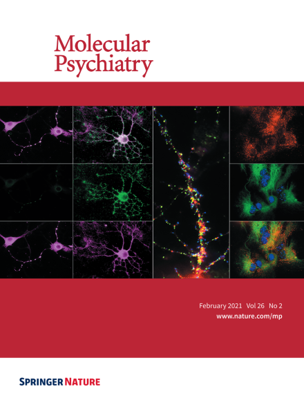
Cell-specific increased surface P2X4mCherryIN in P2X4 internalization-defective knock-in mice. Surface P2X4 (green) is almost absent in MAP2-positive hippocampal neurons (magenta) of control mice expressing wild-type P2X4 but highly increased at the surface of CaMK2Cre-P2X4mCherryIN mice expressing non-internalized P2X4mCherryIN in excitatory neurons (first, second column). Middle, enlarged image of a neuronal dendrite, showing intracellular (red) and surface clustering of P2X4mCherryIN (green) at extra- and post-synaptic regions indicated by Homer 1c (blue). Right column, P2X4mCherryIN (red) is expressed in GFAP-positive (green) astrocytes from hippocampal cultures of CMVCre-P2X4mCherryIN mice. For more information see the article by Eléonore Bertin et al. on pages 629-644.
Image
-
Advertisement
