
Thank you for visiting nature.com. You are using a browser version with limited support for CSS. To obtain the best experience, we recommend you use a more up to date browser (or turn off compatibility mode in Internet Explorer). In the meantime, to ensure continued support, we are displaying the site without styles and JavaScript.

Volume 24 Issue 1, January 2019

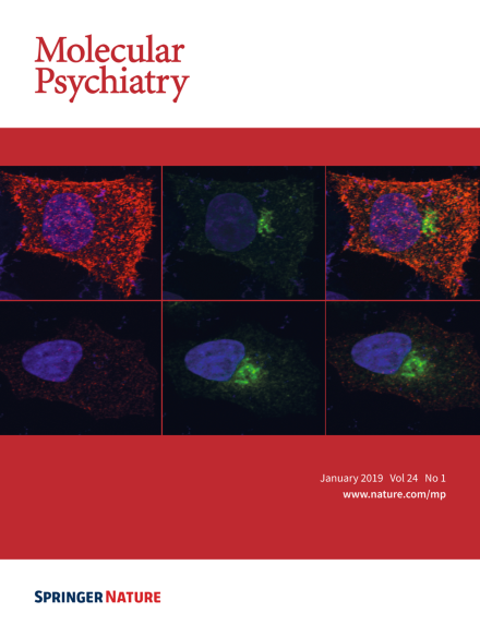
Immunofluorescent labeling of surface (red) and intracellular (green) HA-neuroligin-3 protein in HeLa cells (untreated; top panels) and following the addition of PMA (200 nM for 30 minutes; bottom panel). The activation of PKC dramatically reduces surface neuroligin-3 levels. DAPI was used to label cell nuclei (blue).

For more information, please refer to the paper by Bemben et al. on pages 145–160.

Image

Top of page ⤴

Guest Editorial

Top of page ⤴

Review Article

Top of page ⤴

Expert Review

Top of page ⤴

Article

Top of page ⤴

Search

Quick links