
Thank you for visiting nature.com. You are using a browser version with limited support for CSS. To obtain the best experience, we recommend you use a more up to date browser (or turn off compatibility mode in Internet Explorer). In the meantime, to ensure continued support, we are displaying the site without styles and JavaScript.

NB: AS OF JANUARY 1, 2023 THIS JOURNAL IS PUBLISHED WITH ELSEVIER: https://www.sciencedirect.com/journal/mucosal-immunology

Volume 15 Issue 3, May 2022

 This cover image shows the immunostaining of a gastric tissue section after H. pylori infection which is stained for eosinophil-peroxidase in eosinophils (red) and Foxp3 in regulatory T-cells (turquoise), nuclei were counterstained with Hoechst 33342 (blue). The image shows the infi ltration of eosinophils and regulatory T-cells into the H. pylori-infected gastric tissue and the co-localization of the two cell types in the mucosa.

Volume 15 Issue 3

Obituary

Top of page ⤴

Comment

Top of page ⤴

Review Article

Top of page ⤴

Article

Top of page ⤴

Brief Communication

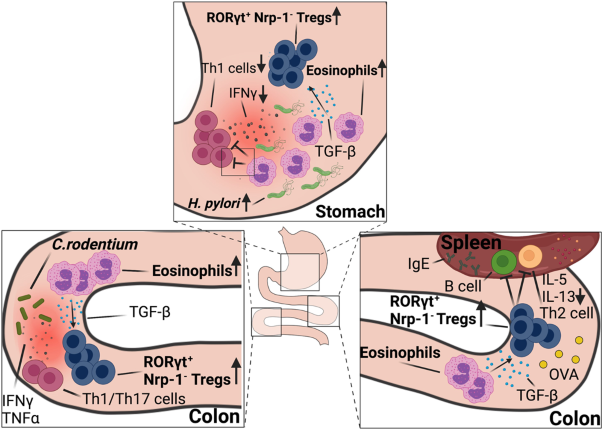
  • Eosinophils are best known for their effector functions in settings of parasitic infection or allergen challenge, but have also increasingly been implicated in immune regulation at mucosal sites. Here, we show using bacterial infection and antigen challenge models that extrathymic Foxp3+ regulatory T-cells that arise de novo in the context of bacterial infection require an intact eosinophil compartment. Mouse strains with a constitutive or conditional eosinophil deficiency, or with an eosinophil-specific ablation of Tgfb, lack bacterially induced neuropilin-negative, RORγt-positive gastrointestinal Treg populations in models of Helicobacter pylori, Helicobacter hepaticus and Citrobacter rodentium infection, as well as in the steady state colon and upon oral ovalbumin challenge. Treg priming in lymph nodes appears not to be impaired. Eosinophil-dependent tissue-resident Tregs express CTLA4, ICOS, CD39 and T-bet in addition to RORγt. Eosinophils reside in close proximity to Tregs in infected tissues, and specifically induce the expansion of newly formed Tregs, but not conventional T-cells in vivo and in vitro. TGF-β expression in eosinophils is induced by bacterial contact and during allergen exposure. Specific Tgfb ablation in eosinophils and the associated Treg defects result in excessive T-cell responses in the examined Th2- but not Th1-polarized settings.

    • Angela Fallegger
    • Martina Priola
    • Anne Müller
    Brief Communication Open Access
  • Monocyte-derived macrophages (MDM) drive the inflammatory response to severe acute respiratory syndrome coronavirus 2 (SARS-CoV-2) and they are a major source of eicosanoids in airway inflammation. Here we report that MDM from SARS-CoV-2-infected individuals with mild disease show an inflammatory transcriptional and metabolic imprint that lasts for at least 5 months after SARS-CoV-2 infection. MDM from convalescent SARS-CoV-2-infected individuals showed a downregulation of pro-resolving factors and an increased production of pro-inflammatory eicosanoids, particularly 5-lipoxygenase-derived leukotrienes. Leukotriene synthesis was further enhanced by glucocorticoids and remained elevated at 3–5 months, but had returned to baseline at 12 months post SARS-CoV-2 infection. Stimulation with SARS-CoV-2 spike protein or LPS triggered exaggerated prostanoid-, type I IFN-, and chemokine responses in post COVID-19 MDM. Thus, SARS-CoV-2 infection leaves an inflammatory imprint in the monocyte/ macrophage compartment that drives aberrant macrophage effector functions and eicosanoid metabolism, resulting in long-term immune aberrations in patients recovering from mild COVID-19.

    • Sina Bohnacker
    • Franziska Hartung
    • Julia Esser-von Bieren
    Brief Communication Open Access
Top of page ⤴

Correspondence

Top of page ⤴

Correction

Top of page ⤴

Search

Quick links