Volume 14
-
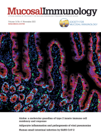
No. 6 November 2021
This cover image shows multiplex IHC imaging of duodenal biopsies from SARSCoV-2 infected individuals stained with antibodies against CD8α (green), EpCAM (red) and Ki67 (cyan). Nuclei were counterstained with DAPI (blue). The image indicates the histomorphological changes in the small intestinal epithelium of COVID-19 patients, characterized by an accumulation of intraepithelial CD8+ T cells, and regenerative proliferation subsequent to epithelial apoptosis. Please see the article in this issue, pages 1381–1392.
-
No. 5 September 2021
This cover image shows 3D volume renderings of 18F-FDG PET/CT scans of a Mycobacterium tuberculosis infected rhesus macaques treated with PBS (above) or the MAIT cell TCR agonist 5-OP-RU (below) from prior to infection, 5- and 10-weeks post-infection (left to right). Image courtesy of Dr. Shunsuke Sakai and Dr. Daniel L. Barber, NIAID/NIH. please see the article in this issue, pages 1055–1066.
-
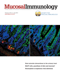
No. 4 July 2021
This cover image shows histologic sections of the small intestine of wildtype (left) and Emc3-deficient mice that was stained with Lysozyme (red), E-cadherin (green) and DAPI (blue). The image indicates the Paneth cells at the bottom of intestinal crypts. Upon Emc3 deletion, these Paneth cells were depleted from intestinal crypts, suggesting Emc3 maintains intestinal homeostasis by preserving Paneth lineage. Image courtesy of Dr. Bing Zhao and Dr. Xinhua Lin, Fudan University. For further information, please see the article in this issue, pages 873-886.
-
No. 3 May 2021
The image shows the expression of protease ELA2A (green), its inhibitor ELAFIN (red) and nuclei (dapi, white) in epithelial cells from human colon biopsies. ELA2A was shown to be secreted by intestinal epithelial cells, where its hyper-activity contributes to intestinal inflammation. Photo courtesy of Anissa Edir and Céline Deraison, INSERM IRSD, Toulouse, France. For more information, please see manuscript in this issue, page 669.
-
No. 2 March 2021
This cover image depicts in situ proximal ligation assay (PLA) using antibodies against CD47 and CD11b on non-permeabilized murine bone marrow-derived neutrophils. Nuclei were counterstained with Hoescht 33342 (blue). This micrograph demonstrates CD47 and CD11b are in close proximity (within 40 nm) and presumably physically associated on the plasma membrane of neutrophils (white dots). Image courtesy of Veronica Azcutia and colleagues, University of Michigan School of Medicine. For further information, please see article in this issue, page 331.
-
No. 1 January 2021
This micrograph shows iron accumulation in the lung of a Mycobacterium tuberculosis - infected C57BL/6 mouse. Iron staining (dark brown) is seen in areas of inflammatory cell infiltration and was markedly decreased in the lungs of mice treated with a pharmacological inhibitor of heme oxygenase-1 compared to those of non-treated animals. The lower intracellular iron accumulation in inhibitor treated mice was associated with increased NOS2 expression and NO production by infected macrophages in response to IFNγ activation as well enhanced bacterial control. For further information, please see the article published in this issue, pp. 253.