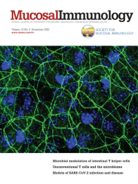
Volume 13
-
No. 6 November 2020
This cover image shows immunofluorescence staining of tight junction protein Claudin-1 in a whole mount cornea from ST2 (IL-33 receptor)-/- mice with experimental allergic conjunctivitis induced by topical eye challenge after sensitization with short ragweed pollen. The image demonstrates that ST2-defi cient mice lack the corneal barrier disruption seen in wild type mice following pollen challenge. Image courtesy of De-Quan Li, Baylor College of Medicine. For further information please see article in this this issue, pages 919–930.
-
No. 5 September 2020
This cover image shows immunofluorescence staining of epithelial sheets of 13 months-old C57BL/6 mice with mAb directed against langerin (green), MHCII (red) and with DAPI (blue). The images demonstrate the distinct morphology of Langerhans cells in aged mice. Image courtesy of Rana Salameh and colleagues, The Hebrew University. For more information please see article in this issue, pp. 767–776.
-
No. 4 July 2020
This cover image shows a histologic section of the urinary bladder that was stained with DAPI (blue), Epcam-1 (white) and F4/80 (red). The image indicates the dense network of macrophages in the lamina propria. Upon infections with uropathogenic E.coli, these macrophages migrate towards the infection to fight off the infection. Image courtesy of Jenny Bottek, Julia Volke and Daniel R. Engel, Department of Immunodynamics, University Duisburg-Essen, Germany. For further information please see article in this issue, page 702.
-
No. 3 May 2020
Salmonella Typhimurium (S. Tm, red) attempts to overcome the intestinal epithelium (wall) to invade systemic body sites (city) with the help of its flagellum. Intestinal epithelial cells (IECs, bricks) and dendritic cells (light blue cells, “police men”) are equipped with NAIP receptors (blue arms) that are able to bind to the flagellum. IECs are highly effective in preventing S. Tm invasion of systemic sites by NAIP-mediated elimination of S. Tm from the intestinal epithelium. S. Tm downregulates its flagellum upon successful invasion (top of the wall) and thereby evades immune cell mediated recognition via NAIP/NLRC4. Illustration by Federico Cecere. For further information please see article in this issue, pages 530-544.
-
No. 2 March 2020
These cover images show histologic sections from the small intestine of CD45.2 mice that received CD45.1 NK cells. The left image depicts an uninfected mouse, while the right depicts a mouse 4 days following infection with Heligmosomoides polygyrus bakeri. The images demonstrate the recruitment of circulatory NK cells in the vicinity of the granuloma following infection. Image courtesy of Maria Elena Gentile and Irah King, McGill University. For further information please see article in this issue, pages 357–370.
-
No. 1 January 2020
Electron microscopy images of the colon of wild type (left), and Lypd8-/- (right) mice infected with C. rodentium. The images demonstrate increased numbers of A/E lesions and internalization of bacteria in the colonic epithelia of Lypd8-/- mice. Image courtesy of Ryu Okumura and Kiyoshi Takeda, Osaka University. For further information, please see the article in this issue, pages 75–85.