Thank you for visiting nature.com. You are using a browser version with limited support for CSS. To obtain
the best experience, we recommend you use a more up to date browser (or turn off compatibility mode in
Internet Explorer). In the meantime, to ensure continued support, we are displaying the site without styles
and JavaScript.
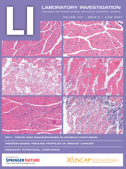
The cover shows morphological manifestations of skeletal muscle contusion in wild-type mice and PD-1-/- mice. For more information, see the paper by Shou et al, this issue (p 719).
Human corneal keratocytes (HCK) are essential for maintaining corneal structure and transparency. This study shows that the natural antioxidant L-carnitine inhibits stromal scarring by suppressing injury-induced intrinsic transient receptor potential vanilloid type 1 (TRPV1) activity that is linked to induction of myofibroblast transdifferentiation in HCK cells. Blocking TRPV1 activation on keratocytes may therefore be a viable approach to suppress corneal opacification in a clinical setting.
Tenascin X-deficient mice exhibit impaired epithelial closure with accelerated infiltration of neutrophils into the tissue and an increased oxidative stress, when compared to wild-type mice. The impairment of epithelial healing in tenascin X-deficient mice was rescued by targeting neutrophil circulation and reducing reactive oxygen species using a chemical scavenger.
The authors examined late- and early-onset murine retinoblastoma tumors and noted that aggressive tumors uniquely expressed an immune gene expression signature. This led to accumulation of a variety of immune cells, including T-lymphocytes, that was not observed in less aggressive tumors. Gene expression analysis identified the immune signature in human and other murine tumors, and they observed it correlated with less proliferative tumors.
Immune cells are involved in skeletal muscle regeneration, but the mechanisms are unclear. In this paper, the authors show that programmed death-1 (PD-1), a key immunosuppressive receptor that is involved in adaptive immune responses, can promote contused skeletal muscle regeneration by regulating Treg cell generation and macrophage polarization.
There exists a void in understanding the communication between classical and nonclassical endocrine signaling. Although progesterone receptor membrane component 1 (PGRMC1) is able to bind to progesterone, the exact signaling mechanism remains unknown. The authors demonstrate that a classical and nonclassical endocrine signaling crosstalk between the estrogen receptor and PGRMC1 exists, and that this crosstalk may promote the growth of breast cancers.
Genome-wide association studies have linked Forkhead Box F1 (FOXF1) to Barrett’s esophagus (BE) but functional data are lacking. Using in vitro models and human material, the authors found that FOXF1 promotes columnar phenotype and cell motility in esophageal squamous epithelial cells, which may have a critical role in BE development.
IL-37 protein is downregulated in endometrial carcinoma cells compared to control endometrium. A mature form of IL-37b (IL-37bΔ1-45) effectively suppresses the migration and invasion of endometrial cancer cells by decreasing the expression of matrix metalloproteinase 2 (MMP2) via the Rac1/NF-κB signal pathway. This results in attenuated tumor metastasis in a peritoneal metastatic xenograft model of endometrial cancer.
The m6A modification level of LncRNA FENDRR is elevated in endometrioid endometrial carcinoma (EEC) cells and the abundant m6A modification promotes FENDRR degradation via recruiting the m6A binding protein YTHDF2. Subsequently, the downregulation of FENDRR resulted in the accumulation of SOX4 protein, thus boosting the proliferation of EEC cells
The breast cancer immune microenvironment was analyzed with the nanoString GeoMx® Digital Spatial Profiler (DSP) in cases from the Carolina Breast Cancer Study. Basal-like breast cancers showed increased expression of markers for regulatory T cells. The results were highly reproducible between whole sections and tissue microarrays.
The authors extensively defined feline low-grade intestinal T-cell lymphoma on the clinical, laboratory, radiological, histologic and molecular levels and demonstrated it is a relevant model for human T-cell lymphoproliferative disorder of the gastrointestinal tract, a rare and poorly described entity. Based on these findings, the group also proposed a new model of digestive T-cell lymphomagenesis.