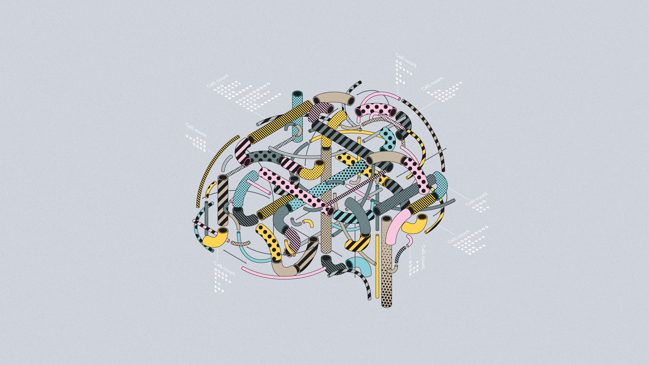
BRAIN Initiative Cell Census Network—Motor Cortex
Generating a multimodal cell census and atlas of primary motor cortex through collaborative data collection, tool development and analysis.

Four years ago, the NIH’s Brain Research through Advancing Innovative Neurotechnologies (BRAIN) Initiative Cell Census Network (BICCN) was launched, with the aim of identifying and cataloguing the diverse cell types in human, monkey and mouse brain. The first instalment of this ambitious endeavour is now complete, with the comprehensive mapping of mammalian primary motor cortical cell type identities on a molecular level.
This collaborative effort has integrated a variety of different large-scale data sets to better define brain cell types, analysing single-cell transcriptomes, chromatin accessibility, DNA methylomes, morphological characteristics and electrophysiological properties in combination with precise anatomical locations.
While the multimodal reference atlas should facilitate the study of brain function by providing detailed data on the ‘parts list’ of motor cortex, this effort from the BICCN has also accelerated the development of a novel toolkit to provide genetic access to various neuronal subtypes, as well as a suite of powerful analytical options for researchers to examine the publicly available datasets for further discovery.
The flagship paper of this BICCN project, published in Nature, aims to establish a unified framework of motor cortical cell-type organization by describing how the various molecular, wiring, and functional components were brought together. Additionally, the flagship serves to validate the BICCN’s systematic strategy of defining cell types, opening up future directions for the application of a similar collaborative and comprehensive approach to generate a brain-wide cell census and atlas of the mouse brain. We will highlight a sample of papers from the package throughout this immersive web feature.
BICCN by the numbers

The BICCN consortium package showcased here includes 17 new articles in Nature and an additional 10 publications across the Nature Portfolio. These research articles cover 136 journal pages in Nature, with 533 total figure subpanels and 258 authors on the flagship paper alone. Like other large-scale consortia, it may take only a few years for the number of publications that use BICCN data to dwarf those published by the BICCN members themselves.
Sample numbers
From just the flagship BICCN publication and its 11 direct companion papers, more than 2.2 million cells or nuclei were analysed for transcriptional information, with more than a million more assessed for chromatin information. Over 1,700 neurons were reconstructed for morphology and projection information, while another 500+ neurons were subject to Patch-seq techniques, collecting molecular, anatomical and physiological information from the same cells.
Data bytes
As of 28 September 2021, the Neuroscience Multi-Omic (NeMO) archive contains 850,292 BICCN files with molecular data deposited for public consumption. These files require 241.3 Tb of storage and vary depending on the type of assay considered. Users have downloaded 478.3 Tb of information from the archive, demonstrating its broad use by the community.

Data courtesy of NeMO Archive.
Data courtesy of NeMO Archive.
BICCN methods and generated tools


Courtesy of Meng Zhang and Xiaowei Zhuang (Harvard University and HHMI).
Courtesy of Meng Zhang and Xiaowei Zhuang (Harvard University and HHMI).
Single cell datasets
Defining the molecular taxonomy of cell types in the motor cortex is best accomplished by examining expression data from cells one at a time. The BICCN flagship initially derived their cell identity definitions from nine datasets, including seven single-cell RNA sequencing collections and two single-cell characterizations of epigenomic features. Further integration of spatially resolved single-cell data using MERFISH provided confirmation and refining of the cell type clusters.
By conducting similar analyses across marmoset and human samples, comparative datasets identified cell classes that were conserved across the mammalian species, as well as divergent subtypes in the cell census, suggesting potential contributions to the differences in brain anatomy and function between the species.
Multimodal data collection from neurons
Patch-seq is an extremely powerful tool that allows the collection of several different datasets from the same cell, allowing the intersection of molecular and functional phenotypes to assist in the refinement of cell type identities. Individual neurons were patched to evaluate their electrophysiological properties, while also being filled with dye to visualize morphological features of the cell and its neurites. At the end of the functional data collection, the nucleus was aspirated for molecular analysis. Not only does Patch-seq allow the comparison of morpho-electric phenotypes with transcriptional data between cells, but it also provides a means to compare potentially related cell types across species and allows proper classification of homologous cells.

Courtesy of Brian Lee and Ed Lein (Allen Institute)
Courtesy of Brian Lee and Ed Lein (Allen Institute)


Cell type targeting tools
Genetic access to specific types of neuron is a critical need for a variety of different experiments, including molecular validation and identification, fate-mapping of progenitors during development, the specific study of neuronal function and more. By splitting excitatory pyramidal neurons on the basis of developmental genetic programs, this resource has created 15 Cre- and Flp- drivers to target glutamatergic neurons of choice, defined by various types of molecular and anatomical data.
By providing improved specificity and coverage of potential pyramidal neuron organization, these tools will enhance the identification and study of biologically important populations of neurons. Combining these tools with established temporal and inducible features will further experimental flexibility in cell targeting, manipulation and fate-mapping. All new knock-in mouse lines will be deposited at Jackson Laboratories, ensuring their wide distribution.
Images courtesy of Hongkui Zeng (Allen Institute).
BICCN results and data

This section showcases a few of the top-line findings from the BICCN papers, which were made possible only through the integration of all the complementary datasets into a single focus on identifying the cell types of mammalian motor cortex.
The scale, depth of data collection and phylogenetic comparisons offered have returned important biological insights well beyond those of a simple atlas or census. The project represents a major advance in our understanding of structure–function relationships in the mammalian brain and should drive innovation in future studies across all areas of neuroscience.
Identifying and defining cell types
By combining and analysing single-cell-derived transcriptional and epigenonomic data, the researchers defined a unified population of cortical cell types in the mammalian motor cortex. Cluster analysis of these integrated datasets yielded more than 50 identified neuronal subtypes. Strikingly, cluster analysis from MERFISH-derived data that included spatial information for each of the cells yielded about the same number of transcriptomic cell types.
Using Patch-seq data, the morphological and electrical properties of the neuron were derived for each transcriptomic subclass, with transcriptionally similar subclasses sharing similar morpho-electric properties.

Courtesy of Meng Zhang and Xiaowei Zhuang (Harvard University and HHMI).
Courtesy of Meng Zhang and Xiaowei Zhuang (Harvard University and HHMI).


Comparative analysis across species
Using similar combinatorial and integrative analyses as for the mouse, additional datasets from marmoset and human brains revealed significant differences in the number of neuronal types in motor cortex; there were 94 clusters in marmoset brain, whereas human samples yielded 127 types. Forty-five transcriptomic types were conserved across all three species, with the most conservation amongst GABAergic neurons. Interestingly, there was substantial evolutionary divergence of the marker genes identified in the cell classes, suggesting adaptations in each of the species or relaxed constraints on molecular function within these neurons.
Images courtesy of the Cytosplore Development Team.
Generating a wiring diagram for motor cortex
A comprehensive, cellular-resolution input–output wiring diagram was established using a variety of methods, including classic tracers, viral labelling and brain-wide imaging. Projections from motor cortex neurons do not seem to follow a simple pattern, with each cell type targeting an individual target region in the brain, but rather form a complex pattern in which multiple inputs are received and multiple regions are targeted by each cell type. As an example, upper limb regions of motor cortex project to more than 100 brain and spinal cord regions, while receiving projections back from around 60 brain areas.
The full axonal reconstructions of 300 neurons revealed a complex range of anatomical types and a diverse relationship with transcriptional cell types. Thus, cell specificity may be refined by additional levels of regulation to determine how a neuron resides in and connects with the cortical network.

Courtesy of Houri Hintiryan and Hong-Wei Dong (UCLA).
Courtesy of Houri Hintiryan and Hong-Wei Dong (UCLA).

courtesy of Hongkui Zeng (Allen Institute).
courtesy of Hongkui Zeng (Allen Institute).
Data access and analytical portals
Another important component of the BICCN is a commitment to rapid dissemination of the data, tools, resources and pipeline structures supported by the project. All data that meet established quality control standards are made publicly available through appropriate data archives. In addition, as knowledge of the relationships between various dataset types becomes more clear, an updated summary of cell-type definitions will be presented.
Transcriptomic and epigenomic data are available through the NeMO archive. NeMO data will include genomic regions associated with disease, transcription activity and regulatory elements, levels of cysteine modification and chromatin accessibility profiles.
Image modality datasets are available through the Brain Image Library (BIL). Here, researchers may analyse, mine or share large brain image resources, as well as using a computational enclave to process datasets.
Neurophysiology data will be archived at the Distributed Archives for Neurophysiology Data Integration (DANDI). DANDI provides a web platform on which researchers can collaborate and process data from physiological recordings, following community data standards. Both electrical and optical neurophysiology recordings are deposited here.
And finally, the Brain Cell Data Center (BCDC), the primary data organizing effort of the BICCN, has generated a comprehensive data catalog, providing access to all of the major datasets.
Browse the collection
View the BICCN collection page which includes all research articles, an editorial, feature article and News & Views.
