Tardigrade circus and a tree of life — January’s best science images
The month’s sharpest science shots, selected by Nature’s photo team.


Tree trails. While taking aerial photos with his drone, Australian photographer Derry Moroney came across these massive, tree-like patterns in Lake Cakora in New South Wales. The colourful drainage channels form when water full of oil from the surrounding tea trees (Melaleuca alternifolia) runs into the lake. The photos were taken after several days of storms. “When I first saw it, I thought it was a tree of life,” Moroney told BBC News. “Every couple of weeks, when we have different weather, it totally changes.”


Special delivery. The Cygnus NG-14 spacecraft just after its release from the International Space Station (ISS), where it had completed the second-heaviest cargo-resupply mission so far. Cygnus carried more than 3,500 kilograms of crew supplies and research equipment to the ISS, including instruments to measure plasma in Earth’s ionosphere and to study the behaviour of fire and smoke in microgravity. The delivery also included a US$23-million, titanium upgraded space toilet for astronauts to test before a similar model is fitted in NASA’s Orion crew capsule for a trip to the Moon.


AI art. Meet DALL-E: an artificial-intelligence (AI) program that can draw pretty much anything you ask for. When asked to draw an armchair in the shape of a pig, this is what the model came up with. DALL-E — named after the Spanish artist Salvador Dalí and the Pixar robot character WALL-E — is a neural network trained on more than 12 billion online images and their captions. It was designed by the company OpenAI in San Francisco, California, to generate pictures when given a short text description (such as “a cat wearing sunglasses” or even “an illustration of a baby daikon radish in a tutu walking a dog”).


Credit: Jes Aznar/Getty
Credit: Jes Aznar/Getty
Broken ground. In January 2020, the Taal volcano in Batangas, the Philippines, erupted for the first time in 43 years, causing earthquakes, lava flows and plumes of ash that blanketed the surrounding area, destroying homes and causing widespread environmental devastation. In the aftermath, traditional sources of income such as fishing have become unviable, and people still cannot return to the island at the centre of the eruption. Here, a scientist collects soil samples at Taal’s summit one year on, as part of an expedition to search for clues on how to make the area productive again.

Credit: S. Gadadhar et al./Science
Swimming laps. These computer-enhanced microscope images show how genetic mutations that affect the microstructure of sperm’s flagella, or tails, can affect the cells’ swimming ability. The top sperm follows a normal, linear swimming path, shown by the coloured trails. The middle and bottom sperm carry mutations that affect the structure of their tails, causing them to swim in circles, or diagonally. Both lack enzymes that make important modifications to a protein called tubulin — a major component of the tail core. The findings point to a mechanism underlying certain types of infertility.

Credit: HEPIA
Miniature mind. This is a seven-month-old ‘mini-brain’ — a pinhead-sized ball of different types of human brain cell — seen under a confocal microscope. Scientists use mini-brains to study brain development and disease progression. To look inside one, researchers usually have to first cut it into thin slices. But a team at the Geneva School of Engineering, Architecture and Landscape (HEPIA) and the Wyss Center for Bio and Neuroengineering in Geneva, Switzerland, has developed a technique that allows it to produce 3D images of intact mini-brains, revealing the structure and positioning of individual neurons in detail. “Despite advances in growing mini-brains, it has been difficult to understand in detail what is going on inside — until now,” says Adrien Roux, a bioengineer at HEPIA.
Credit: David Prötzel
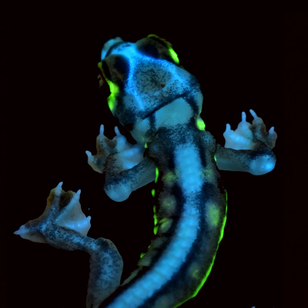
Glowing gecko. This baby Namib web-footed gecko (Pachydactylus rangei) fluoresces under ultraviolet light. The intense neon-green and blue glow — among the brightest fluorescence in any vertebrate — is produced by modified pigment cells called iridophores. Why many animals fluoresce is still a mystery — but in this case, the pattern suggests that it helps these social animals to signal to each other across the moonlit desert.

Credit: S. Gadadhar et al./Science
Credit: S. Gadadhar et al./Science


Credit: HEPIA
Credit: HEPIA


Credit: David Prötzel
Credit: David Prötzel
Credit: Penelope Fenton. This video has no sound.
Credit: Penelope Fenton. This video has no sound.
Tardigrade wheels. Tardigrades, also called water bears, are best known for being indestructible — they can survive extreme heat, radiation and even the vacuum of outer space. This video of a tardigrade under the microscope shows how it interacts with spherical colonies of Volvox aureus, a green alga. “Tardigrades really like something to hold on to when they are on a slippery glass slide, so naturally they grab on to just about anything they waddle into,” says Penny Fenton, a PhD student at the University of Suffolk in Ipswich, UK, who made the video. “They do eat algae, but the Volvox are safe as long as they keep moving.” Fenton studies aquatic toxicology, and does microphotography in her spare time. She cultures the algae and finds wild samples of tardigrades in leaf litter and water from her shed’s rain gutters. Videos of her microscopic performers on Instagram have attracted thousands of likes.
Credit: Penelope Fenton. This video has no sound.
Credit: Penelope Fenton. This video has no sound.
Tardigrade wheels. Tardigrades, also called water bears, are best known for being indestructible — they can survive extreme heat, radiation and even the vacuum of outer space. This video of a tardigrade under the microscope shows how it interacts with spherical colonies of Volvox aureus, a green alga. “Tardigrades really like something to hold on to when they are on a slippery glass slide, so naturally they grab on to just about anything they waddle into,” says Penny Fenton, a PhD student at the University of Suffolk in Ipswich, UK, who made the video. “They do eat algae, but the Volvox are safe as long as they keep moving.” Fenton studies aquatic toxicology, and does microphotography in her spare time. She cultures the algae and finds wild samples of tardigrades in leaf litter and water from her shed’s rain gutters. Videos of her microscopic performers on Instagram have attracted thousands of likes.
Crown of thorns. Visual representations of the coronavirus SARS-CoV-2 have become a common sight over the past year. But this particular image shows real coronavirus particles in unprecedented detail. It is the first 3D image of SARS-CoV-2 made from a single scan of frozen virus particles, using a technique called cryo-electron tomography. Most previous images were either composites from several scans, or computer visualizations. Researchers at the Vienna-based company Nanographics added colours to distinguish different parts of the virus.