

Key Points
- Many disorders of the central nervous system (CNS) can cause oral and pharyngeal dysphagia.
- For clinical purposes, the disorders can be classified as nondegenerative or degenerative. Degenerative disorders are further subclassified based on their main clinical presentation into dementia, movement disorders, and others.
- Stroke is the most common type of nondegenerative disorder. Degenerative disorders are usually progressive.
- Functional swallowing abnormalities include disordered preparatory phase, poor bolus control, difficulty initiating a swallow response, and decreased hyolaryngeal elevation. Aspiration is the most clinically significant consequence of dysphagia.
- The swallowing abnormalities in CNS disorders are generally not specific to the type of neurologic disorder.
- Diagnosis of swallowing dysfunction requires careful history and clinical examination, but videofluoroscopy is the gold standard for characterizing the specific abnormality. Videoendoscopy may also be useful in certain circumstances.
- Videofluoroscopy and videoendoscopy are also valuable in identifying and teaching maneuvers that may facilitate swallowing and prevent aspiration in a patient.
- When significant aspiration cannot be prevented, alternatives to oral feeding such as percutaneous endoscopic gastrostomy (PEG) tube placement should be considered.
- Patients with oropharyngeal dysphagia owing to CNS lesions are best managed by a team approach including a speech pathologist, neurologist, and gastroenterologist.
Introduction
Neurologic disorders affecting swallowing can be categorized in many different ways, including by the anatomic site of the lesion such as the central or peripheral nervous system, by the pathologic process such as ischemia and degeneration, by the underlying etiology, or by the clinical presentation such as dementia and movement disorders. Often several of these parameters are used in a classification scheme (Table 1). Disorders of the central nervous system (CNS) can be nondegenerative or degenerative. Based on the clinical progression, degenerative disorders may be subclassified into progressive and relapsing disorders. Progressive degenerative disorders may be further subclassified based on their salient clinical characteristic into dementias and movement disorders. In contrast to the CNS, most of the peripheral nervous system disorders that impact swallowing are degenerative in nature.1

Stroke
Stroke affects 2000 people per million worldwide each year2 and approximately 700,000 individuals in the United States annually.3 Eighty percent of strokes are secondary to ischemia, which results from internal carotid artery atherosclerosis or cardiac embolism, whereas hemorrhage accounts for 20%. Neurologic symptoms of stroke vary depending on the specific location of stroke. With a supratentorial infarct, symptoms may include ipsilesional hemiparesis, dysarthria, aphasia, and hemispatial neglect. With a lateral medullary infarct, neurologic presentation may include ataxia, reduced ipsilesional limb pain and temperature sensation, reduced contralesional pain and sensation of the trunk and limbs, and ipsilesional velar, pharyngeal, and laryngeal paralysis. Swallowing abnormalities in stroke are variable and may include oral lateral sulci retention, delayed oral transfer, delayed elicitation of a pharyngeal swallow, decreased hyolaryngeal elevation, and aspiration.4
Traumatic Brain Injury
Traumatic brain injury can result in dysphagia depending on the brain region involved. Brain injury resulting from trauma is generally more diffuse than that following stroke, and cognitive impairments are often prominent depending on the site and severity of injury.
Brain Tumor
As with stroke and traumatic brain injury, brain tumors may result in dysphagia depending on which brain region is involved. Pathologically, brain tumors may be benign or malignant; however, their space-occupying nature can result in significant neurologic dysfunction. Swallowing may be affected by infiltration of the tumor into brain regions important in deglutition. In addition, the treatment modalities for the tumor including surgery and radiation therapy may affect swallowing. Unlike the dysphagia resulting from stroke or traumatic brain injury, swallowing disorders secondary to a brain tumor may be progressive as the tumor invasion increases.
Cerebral Palsy
Cerebral palsy results from brain damage in utero or at birth and yields chronic neurologic problems. Severity of deficits is dependent on the severity of the brain damage.
Iatrogenic
Medication Induced
Tardive dyskinesia is the most common type of medication-induced disorder and results from the chronic dopamine-receptor blockade that is generally associated with neuroleptic drugs. It can occur in up 20% of patients receiving neuroleptics. Neurologic symptoms include repetitive movement of the lower face, lips, and tongue (orobuccolingual dyskinesia), and movement of the trunk and pelvis. Symptoms may be reversible if identified early and the medication changed to newer medications such as Risperdal, which does not produce these symptoms.
Surgery Induced
Surgery involving the neck, such as cervical spine surgery and carotid endarterectomy, may produce dysphagia generally owing to manipulation of cranial nerves. The anterior approach frequently used with cervical fusion results in dysphagia more frequently than a posterior approach.5 Dysphagia, which often co-occurs with postoperative dysphonia, results from retraction and stretch injury of cranial nerves. Generally damage to the pharyngeal plexus may occur with anterior cervical fusion. Injury of the seventh, tenth, and twelfth cranial nerves may occur with carotid endarterectomy, as these nerves are close to the carotid bifurcation. Postoperative edema may also contribute to the dysphagia.
Dementia
Dementia is characterized by a decline in one or more major cognitive domains (i.e., language, visuospatial functions) accompanied by impairment in memory. There are multiple causes of dementia. Patients with dementia not only have problems with swallowing but also with feeding in that they may have limb apraxia affecting their ability to use eating utensils and agnosia affecting their ability to recognize and accept food. Deficits in transfer of the bolus in the oral cavity are also prominent.4
Alzheimer's disease (AD) is the most common type of dementia. Neuritic plaques and neurofibrillary tangles are the neuropathologic hallmarks of AD and result in severe brain atrophy with widened sulci and gyri shrinkage. Alzheimer's disease is generally sporadic, but a familial autosomal dominant variant has been identified. Initial symptoms include decreased recent memory and anomia. As the disease progresses, symptoms include inability to complete activities of daily living, agitation, and mutism. Age is the major risk factor of AD with occurrence increasing exponentially after the age of 65 years; however, in the familial variant, onset is earlier.
Frontotemporal dementia (FTD) is associated with severe atrophy of the frontal and temporal lobes with prominent changes in behavior. As with AD, the occurrence of FTD is primarily sporadic, but familial occurrence due primarily to the tau gene mutation is also evident. Frontotemporal dementia may co-occur with amyotrophic lateral sclerosis, which is discussed later. Symptoms of FTD include personality changes, mental rigidity, decreased language, stereotyped behavior, hyperorality, and decreased ability to perform activities of daily living.
Lewy body dementia, as with AD and FTD, is typically sporadic but may also be familial. Many patients with Lewy body dementia have first-degree relatives with Parkinson's disease or dementia. Neurologic characteristics include fluctuating cognitive ability, visual hallucinations, spontaneous parkinsonism, sleep disturbance, and reduced ability to perform activities of daily living.
Vascular dementia may result from multiple cortical or lacunar infarcts, a single strategically placed infarct, or microvascular disease. Characteristics include a slow, stepwise, and fluctuating progression with memory frequently mildly affected. Dysfunction in executive control is evident early and is characterized by disorganized thought, behavior, and emotion. Gait is frequently disturbed. Vascular risk factors are prominent with infarcts or white matter lesions evident on neuroimaging studies.6
Movement Disorders
Movement disorders constitute a group of degenerative metabolic disorders of the CNS involving the extrapyramidal and cerebellar pathways, which are characterized by disordered movement that allows for their easy clinical recognition.
Parkinson's disease (PD) is the most prevalent of the movement disorders. It is a degenerative disorder involving dopaminergic neuronal loss in the striatal tract of the basal ganglia. Onset of PD typically occurs between the ages of 55 and 65. Onset and progression of symptoms is gradual and initially asymmetric. Neurologic symptoms initially include resting tremor, bradykinesia, and rigidity. As the disease progresses, symptoms may include autonomic disturbances and cognitive and behavioral changes.
Progressive supranuclear palsy (PSP) is a rare, generally sporadic, rapidly progressive disease involving the basal ganglia, frontal lobes, midbrain, and brainstem. Onset typically occurs between the ages of 50 and 60 years. Symptoms include vertical ophthalmoplegia, axial rigidity, pseudobulbar palsy, and dementia.
Olivopontocerebellar atrophy is one of a group of degenerative diseases involving multiple system atrophy, with the atrophy in olivopontocerebellar atrophy concentrated in the cerebellum, pons, and inferior olivary nuclei. The variants of olivopontocerebellar atrophy include genetic (autosomal dominant and recessive) and sporadic forms. Onset of olivopontocerebellar atrophy occurs generally within the sixth decade. Neurologic symptoms include gait disturbance, dysarthria, dysphonia, and dysphagia.
Huntington's disease (HD) is a hereditary degenerative autosomal dominant disease involving a preferential loss of  -aminobutyric acid (GABA)ergic neurons, which yields diffuse atrophy with concentrated cell loss in the caudate and the putamen. Onset occurs between the ages of 30 and 50 years. Neurologic symptoms include gait disturbance, dysarthria, dysphonia, and dysphagia.
Wilson's disease is a rare hereditary degenerative autosomal recessive disease resulting in a disorder of copper metabolism. Symptoms include liver disease, tremor, rigidity, dystonia, ataxia, dysarthria, and psychiatric disturbances. Symptoms of liver disease occur in childhood, with approximately half of Wilson's disease patients presenting with neurologic symptoms during adolescence.
Multiple Sclerosis
Multiple sclerosis is an autoimmune disease caused by an inflammatory demyelinating process that affects multiple white matter tracts within the CNS. Symptom onset is generally between the ages of 25 and 30 years. Clinical features are varied in multiple sclerosis, reflecting the multifocal areas of the CNS and may include bilateral internuclear ophthalmoplegia, sensory impairment, heat sensitivity, and fatigue involvement.
Amyotrophic Lateral Sclerosis
Amyotrophic lateral sclerosis (ALS) is a progressive disorder affecting both the upper and lower motor neurons. Between 5% and 10% of cases are believed to be the result of an autosomal dominant mutation. Onset is generally around the age of 60 but may occur in patients as young as 20 years of age. ALS can affect predominately the corticobulbar or corticospinal tracts. The spinal presentation generally includes reduced dexterity, limb weakness, and spasticity, whereas the bulbar presentation includes dysarthria, dysphonia, dysphagia, sialorrhea, muscle atrophy, and fasciculations. It is generally believed that cognitive functioning is spared; however, formal neuropsychological assessment is often precluded by the patient's loss of speech and motor functions in the later stages of the disease. Some estimates suggest that FTD may occur in approximately 5% of patients with ALS. The disease is always fatal, with death generally occurring within 2 to 5 years of diagnosis of corticobulbar ALS.
Impact of New Technology on the Understanding of Dysphagia in CNS Disorders
Although dysphagia has been reported as a symptom of CNS disease for almost two centuries,7, 8 it was generally believed that bilateral hemispheric or brainstem damage must occur to produce dysphagia. Clinical evaluation and postmortem brain studies9 and cortical stimulation studies in humans,10 however, demonstrated that damage to unilateral cortical brain regions could produce dysphagia. Nonetheless, it was not until the application of neuroimaging techniques that it became widely accepted that dysphagia could result from unilateral supratentorial damage.11, 12 Furthermore, it is now known that a lesion in specific sites can produce dysphagia. These sites include the premotor and primary motor cortices, primary somatosensory cortex, insula, as well as the periventricular white matter, which disrupt cortical-subcortical connectivity when lesioned.13, 14, 15, 16 Functional neuroimaging studies in healthy adults using positron emission tomography and functional magnetic resonance imaging have provided additional insights regarding the neural substrates of swallowing.17, 18, 19, 20
It was only with the advent of cinefluorography that the dynamics of swallowing could be observed in vivo. Routine evaluation of the oropharyngeal swallowing mechanism became possible following the development of this technology. Improved specificity of oropharyngeal dysmotility patterns secondary to neurologic disease has been documented over the past two decades using instrumental evaluation, specifically videofluoroscopic swallow studies (VSSs) and the use of videoendoscopy to evaluate swallowing has provided additional information on deglutitive function. As a result of objective measures of specific biomechanical dysfunction producing dysphagia, evidence-based approaches to the rehabilitation of neurogenic swallowing disorders have become available over the past 15 years.
Epidemiology of Dysphagia in CNS Disorders
It is estimated that 400,000 to 800,000 individuals worldwide develop neurogenic dysphagia per year.21 The reported incidence of dysphagia in specific neurologic diseases is variable, owing in part to patient selection methods (e.g., consecutive patients, case series) and evaluation methods (e.g., questionnaire, clinical evaluation, diagnostic evaluation). Determination of dysphagia based on patient complaint or clinical swallowing evaluation generally underestimates the incidence. It is generally agreed that stroke is the most common cause of neurogenic dysphagia. Dysphagia, as identified by VSSs, occurs in approximately 65% of acute stroke patients,22 with the incidence of aspiration ranging from 43% to 58%.22, 23, 24 Of acute stroke patients who aspirate, 40% to 70% do so silently.22, 25 That is, there is no overt sign of aspiration such as cough or voice change when material contacts the true vocal folds or enters the trachea. See Table 2 for the prevalence of dysphagia in other neurologic disorders.
Functional Characterization of Dysphagia in CNS Disorders
Neurogenic dysphagia can result in dysfunction in oral preparation, oral transfer, and pharyngeal motility. A delayed pharyngeal swallow is an example of a disturbance in the sensory system, whereas reduced tongue control with premature spillage of material into the pharynx is an example of a disturbance in the motor system. Depending on the neurologic disorder, cognition may also be impacted. Cognitive deficits, such as those evident in HD, are associated with feeding abnormalities, which contribute to dysphagia. Agnosia may contribute to the lack of oral transfer as evident in AD.
Clinical Features of Dysphagia in CNS Disorders
Patients with CNS disorders may present clinical signs of swallowing problems. Prandial features may include drooling, reduced mastication, throat clearing, coughing and choking, nasal regurgitation, and complaints of residue in the oral cavity and pharynx. Coughing and choking, however, may not present in patients who aspirate silently. Moreover, reduced sensation or cognition associated with many neurologic disorders may impair a patient's awareness of the symptoms. Weight loss, dehydration, and respiratory compromise may also be associated with dysphagia. Diet alteration to include softer foods and the use of compensatory strategies may be spontaneously implemented by patients with dysphagia to facilitate swallowing.
Other clinical features evident in neurogenic diseases include sialorrhea (hypersalivation), which can be present in diseases such ALS and PD, and eructation (excessive burping), which is characteristic in HD. Although present in neurologic diseases with a high incidence of dysphagia, it is unclear if sialorrhea and eructation are related to disturbed deglutition.26, 27, 28
Gag Reflex as a Marker of Dysphagia in CNS Disorders
For decades, it has been assumed that an absent gag reflex is indicative of neurogenic dysphagia. Although an abnormal gag reflex may be apparent in patients with dysphagia resulting from various neurologic disorders, it may be absent in healthy control subjects or it may be normal in patients with neurogenic dysphagia.29 Thus, when observed in isolation, the integrity of the gag reflex provides no salient information about swallowing, and as such, determination of oral intake should not be based on this sole feature.
Role of Clinical History in the Diagnosis of Dysphagia in CNS Disorders
Careful history intake is critical in the workup of neurogenic dysphagia. The onset of dysphagia and the CNS disorder are frequently related. For example, stroke is associated with a sudden onset of dysphagia, whereas degenerative disorders such as PD are associated with a gradual onset of swallowing problems. Although it is important to determine the onset of dysphagia, many patients with CNS disease may be unaware of their dysphagia. This may be owing to decreased sensation, in which a cough reflex is not evoked with aspiration or there is no awareness of pharyngeal retention. Other factors that may interfere with patient awareness include cognitive deficits and voluntary or involuntary compensatory strategies that reduce obvious symptoms of dysphagia.30 Accordingly, the caregiver may need to be present when obtaining the case history, which should include direct questions regarding swallowing, length of time to complete meals, food avoidance, and weight loss, even if the patient voices no concern with swallowing.
Overt symptoms of oropharyngeal dysphagia may include coughing when eating or drinking, a voice change (particularly "gurgly" vocal quality) during meals, and weight loss. Patients may complain of food sticking in their throat, which may indicate pharyngeal or esophageal retention. Regardless of the symptom, it must be emphasized that the clinical symptoms of neurogenic dysphagia are generally nonspecific and may not correlate with the underlying disease process.
Role of the Clinical Examination in the Evaluation of CNS Dysphagia
The clinical swallowing evaluation generally includes an examination of orofacial muscular symmetry, strength, and sensation, cranial nerve assessment, and evaluation of voice and speech. Swallowing is also assessed using a variety of liquid volumes as well as with substances of various consistencies.
Clinical Identification of Patients at Risk for Dysphagia
The main purpose of the clinical swallowing evaluation is to identify patients who are at risk for dysphagia, thus warranting a diagnostic swallowing evaluation. This is important, particularly following an acute stroke, as all patients may not warrant a diagnostic examination. As such, research has focused on identifying critical clinical features that may help to predict dysphagia and risk of aspiration following a stroke.
Initially, such studies focused only on swallowing to identify the risk of aspiration in stroke regardless of time post-onset. These studies relied on the presence of a cough or voice change following ingestion of three ounces of water31 or the rate of swallowing32 to determine risk of aspiration. Although illustrative, the results of such studies were hampered methodologically by failing to identify patients with silent aspiration, and the self-administration of large volumes of water may have increased the risk of aspiration. Poor sensitivity has confounded the results of some research relying on the 3-oz test, in which numerous patients passed the clinical test but aspirated during the VSS.33
Daniels and colleagues22, 23 studied other factors in addition to results from a clinical swallowing evaluation, and they identified six clinical features associated with the risk of aspiration (i.e., residual laryngeal penetrated material or aspiration) as confirmed by VSS in acute stroke patients: abnormal volitional cough, abnormal gag reflex, dysarthria, dysphonia, cough after trial swallow, and voice change after trial swallow (Table 3). The presence of any two of these six clinical features correctly identified the risk of aspiration with 92% accuracy. Implementation of this clinical pathway was initiated at the Veterans Affairs Medical Center in New Orleans, in which acute stroke patients with two or more of the six clinical features underwent a VSS, and patients with less than two clinical features did not receive the instrumental examination.34 Based on patient outcomes and VSS results, implementation of the clinical pathway distinguished patients with moderate to severe dysphagia from patients with mild dysphagia or normal swallowing. None of the patients developed pneumonia, and most returned to a regular diet. Studies by other investigators have confirmed the utility of these six clinical features in the identification of the risk of aspiration in acute stroke patients but have suggested that the presence of four clinical predictors increases specificity.35 Others have indicated that the presence of two of these six features identified by Daniels et al. was not strongly related to aspiration as identified by endoscopic evaluation36; however, the two studies differed in outcome (definition of aspiration risk), evaluation type, and evaluation protocol (numerous volumes and viscosities in the Daniels study compared to a 5-mL volume in the Leder36 study).
Dysarthria may correlate with dysphagia with bulbar ALS. Patients with this disease may not complain of dysphagia but may evidence reduced speech intelligibility. Strand et al.37 found that the progression of dysphagia parallels the progression of speech intelligibility in ALS. Thus, swallowing should be instrumentally evaluated in patients with ALS who present with dysarthria even though they may be without complaints of dysphagia. Furthermore, these researchers found that dysphagia increases as respiratory capacity decreases regardless of the form of ALS. As such, vital capacity should be consistently measured. Accurate and timely assessment of a clinically relevant decline in respiratory status is crucial for determining the timing of feeding tube placement, as much if not more so than an accurate assessment of swallowing ability.
Bedside Diagnostic Tests
Diagnostic studies have been added to the clinical evaluation of stroke patients in order to increase objectivity.
Periprandial pulse oximetry involves measurement of oxygen saturation before, during, and after swallowing. Contradictory findings have been reported, with some studies indicating a strong correlation between desaturation and aspiration,38, 39 whereas others have shown poor association.40, 41 Such discrepancies may be attributed to nonuniformity of techniques used in the different studies.
Nebulized tartaric acid, a mild chemical irritant, has been used to identify stroke patients at risk of developing pneumonia. This evaluation requires patients to inhale tartaric acid that is administered with a nebulizer. A maximum of three inhalations are administered; testing is terminated after the third inhalation or with evocation of a cough reflex after inhalation. Initial research revealed that none of the patients with a normal cough reflex developed pneumonia, whereas 17% of patients who did not pass the test developed pneumonia.42 It is important to note that correlation with imaging studies of swallowing was not completed in any of the research studies and that medical and therapeutic interventions after clinical testing were not described.
Thus far, the clinical features for the risk of aspiration have not been identified for populations other than acute stroke. Moreover, only one study has attempted to identify features from clinical swallowing examination to determine oral and pharyngeal dysphagia, as well as the risk of aspiration.43 Specificity and sensitivity were generally low for each outcome, possibly owing to the heterogeneous sample, which included patients with neurologic disorders, head and neck cancer, etc. Research should focus on identifying clinical features associated with dysphagia, not just aspiration, on homogeneous neurogenic populations.
Role of Imaging Studies in the Evaluation of CNS Dysphagia
The purpose of imaging examinations is to directly evaluate physiologic functioning of the oropharyngeal swallowing mechanism, determine swallowing safety, and identify the effects of compensatory strategies, such as posture and bolus consistency, on deglutition. By determining the exact cause of dysfunction, therapeutic intervention can be initiated to address the specific disorder. The two imaging tools used to evaluate oropharyngeal dysphagia are VSS and videoendoscopy. Although the advantages and disadvantages of each have been well documented,44, 45 no study has compared the two tests in a homogeneous neurogenic populations; thus it is difficult to determine the superiority of either instrument in the evaluation of specific neurogenic disorders.
As dysphagia owing to CNS disorders can impair all three stages of swallowing, VSS is the only tool that allows for direct assessment of the oral cavity and pharynx and allows for the examination of esophageal functioning. Swallowing functioning can be assessed before, during, and after the swallow. The Penetration-Aspiration Scale46 provides an objective way during the VSS to measure the depth, response, and clearance of material entering the larynx and trachea.
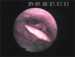
An advantage of the videoendoscopic evaluation is that it can be completed at the patient's bedside or in the office using real food without limitations on the length of the assessment (see Video 1 for a videoendoscopic swallowing evaluation of neurogenic patient). Laryngeal functioning and the ability to control secretions can be assessed. Laryngopharyngeal sensation may also be evaluated with a specially designed videoendoscope that allows for delivery of air-puff stimulation to the medial surface of the pyriform sinuses and aryepiglottic folds in order to evoke a laryngeal adductor reflex.47 Although research has suggested that reduced laryngopharyngeal sensation in stroke patients contributes to dysphagia,48 the association of the laryngeal adductor reflex and response to airway invasion during swallowing has not been determined.
Video 1: Videoclip of VSS in stroke patient showing premature spill.
Note that liquid material enters the pharynx and larynx prior to activation of swallow reflex. White out (screen obliteration) occurs during the actual swallow.
Manometry, although not an imaging study but frequently completed simultaneously with VSS, measures pharyngeal and upper esophageal sphincter (UES) pressures. Pharyngeal manometry allows for measurement of intrabolus pressure, timing of pharyngeal contraction, and relaxation of the cricopharyngeal muscle. Before a surgical procedure, such as cricopharyngeal myotomy, is completed, manometric studies should be completed to assess functioning of the cricopharyngeal muscle during swallowing.
Clinical Laboratory Tests
Laboratory tests may be useful in identifying nutritional deficits caused by dysphagia and may facilitate determination of the underlying neurologic etiology of dysphagia. Screening for vitamin B12 deficiency, thyroid dysfunction, and syphilis must be completed in the workup for Alzheimer's disease in order to rule out potential other causes of dementia. Laboratory studies that are routinely performed in the workup of CNS disorders causing dysphagia include compete blood count, chemistry panel, creatine kinase, vitamin B12, thyroid screening, antiacetylcholine receptor antibodies, and serologic test for syphilis.30
Correlation between Swallowing Abnormalities and CNS Disorders
As is evident from Table 4, there are generally no distinguishing features of oral or pharyngeal stage dysmotility that can differentiate various neurologic diseases. Oral transfer or the pharyngeal swallow may be delayed, and aspiration and residue may be present in almost any given neurologic disorder. There are, however, some exceptions to this rule, and some distinctive patterns that are evident on the instrumental examination may help specify the underlying neurologic disorder. Logemann4 suggests the repetitive "rocking and rolling" festinating-type motion of the tongue during oral transfer is frequently evident in patients with PD. Patients with dementia may have agnosia and hold the bolus in the oral cavity for extremely long periods without attempts to swallow. Patients with HD and FTD frequently demonstrate tachyphagia, in which rapid and unregulated amounts of food and liquid are placed in the oral cavity and food may be swallowed before mastication is complete. In the pharyngeal stage, an extremely weak or absent pharyngeal swallow is generally associated with a lateral medullary stroke. It has been suggested that PD and PSP may be differentiated by silent aspiration, neck extension, and lingual tremors, which may be evident in PD but generally not in PSP,49 whereas others have suggested that the two diseases cannot be distinguished based on the results of the VSS.50
Some studies have suggested differences in oral and pharyngeal functioning based on the hemisphere lesioned in stroke patients. That is, oral stage dysmotility may be associated with left hemispheric damage, whereas pharyngeal stage dysfunction and aspiration may be associated with right hemispheric damage.11, 12 Others have suggested that hemisphere does not distinguish between dysphagia features and aspiration.13, 14 It is generally agreed that swallowing initially may be more severely impaired following a lateral medullary stroke compared to a stroke of the supratentorial region.
Diagnostic Approach to Dysphagia in CNS Disorders
Diagnosis of dysphagia in CNS disease requires two parallel approaches: diagnosis of the CNS disease and determination of the swallowing deficit, particularly identification of aspiration. The swallowing evaluation includes a careful history, clinical evaluation, and imaging study of swallowing. This approach facilitated determination of the severity of the dysphagia, the most appropriate mode of nutritional intake (oral versus nonoral), and the prognosis for recovery of swallowing. The swallowing evaluation does not decipher the nature of the underlying CNS disorder. A careful history, neurologic examination, and specialized tests under the direction of the neurologist are required to determine the neurologic diagnosis.
Clinical Course and Complications of Dysphagia in CNS Disorders
Dysphagia in some neurologic diseases, such as stroke or traumatic brain injury or following neck surgery may demonstrate spontaneous recovery or rapid improvement with therapy within the first few weeks of the event. Patients may warrant initial nonoral intake based on the results of the swallowing imaging study, but should rapidly progress to oral intake within a month following the onset of the neurologic event. It has been suggested that recovery of swallowing in acute stroke patients may be rapid, warranting reassessment within 3 weeks of the initial swallowing evaluation.51 Although most stroke patients may return to an oral diet at 1 month post-stroke, abnormalities in swallowing are still evident on the VSS in most stroke patients.52
Many of the neurologic disorders that affect swallowing are progressive; thus swallowing can be expected to decline as the disease worsens. Some diseases such as multiple sclerosis may progress slowly, whereas other diseases such as ALS, particularly with bulbar presentation, progress rapidly. Swallowing frequently parallels the course of the disease, as seen in patients with ALS, PSP, and HD. For example, a progressive decrease in respiratory capacity in patients with ALS will, in turn, impact swallowing. To complicate matters, the ability to provide surgical intervention to initiate nonoral intake in ALS patients is dependent on the patient's respiratory capacity. By the time patients with ALS require tube feeding, their respiratory capacity may be too impaired to safely tolerate the surgical intervention.37 In its position statement, the American Academy of Neurology recommended a vital capacity of greater than 50% prior to percutaneous endoscopic gastrostomy (PEG) tube placement,53 as research has indicated increased mortality following PEG placement in ALS patients with a vital capacity that falls below this level.54
Language and cognitive impairment may result from both nondegenerative and degenerative neurologic diseases. This can impact recovery as a person may not be able to participate in direct swallowing treatment or may not be able to comprehend or recall specific compensatory strategies. In such cases, it may be necessary to extend the duration of nonoral intake in patients with nondegenerative diseases to maintain safety or adequate nutrition, or to initiate nonoral intake earlier in patients with degenerative diseases to more quickly accommodate the patient and caregivers to a feeding routine that will become even more necessary as the disease process progresses.
Pneumonia can be a frequent complication in patients with dysphagia owing to CNS disease, in part because of unawareness of the swallowing problem and silent aspiration. However, impaired physical and cognitive abilities, which may be frequent in specific neurologic diseases, may exacerbate this problem. In a seminal study, Langmore and colleagues55 identified predictors of pneumonia: dependency in feeding, dependency in oral care, number of decayed teeth, tube feeding, more than one medical diagnosis, number of medications, and presently smoking. Dysphagia and aspiration (of food more so than liquid) were identified as important causes of pneumonia only if these other risk factors were present. Thus, neurogenic patients with cognitive decline or reduced physical ability requiring assistance in activities of daily living may be at greater risk for developing pneumonia.
Management of Dysphagia in CNS Disorders
Direct swallowing rehabilitation, as well as any medical or surgical intervention for dysphagia, should be performed to address a specific swallowing disorder identified with the instrumental examination. Accordingly, the instrumental examination guides the course of treatment. If swallowing dysfunction is evident during the instrumental evaluation, therapeutic intervention must be attempted during the instrumental evaluation in order to assess the efficacy of the intervention relative to the particular deglutitive dysfunction. The employment of a therapeutic strategy must be specific to the actual biomechanical disorder and not merely applied in a random fashion.
Rehabilitation
Generally the first step in the rehabilitation of swallowing deficits is the initiation of swallowing therapy. Most of the research for specific swallowing treatments is composed of case series or single subjects, with stroke being the most common neurologic disorder studied. Before determining the appropriate swallowing rehabilitation program, it is important to understand a patient's cognitive, motor, and sensory abilities, as any or all of these can be adversely affected by neurologic disease and may impact swallowing recovery. For example, utilization of a chin tuck posture and thickened liquids may both decrease the risk of aspiration of liquids for a delayed pharyngeal swallow. However, in a patient with AD or HD, thickened liquids may be more appropriate, as the patient may not remember to implement or cannot maintain a chin tuck posture.
Swallowing therapy may include compensatory or rehabilitative strategies.4, 56 Compensatory therapy does not change the physiology of the swallow; rather, bolus flow is redirected. Compensatory strategies are attempted during the instrumental study to determine their effectiveness. These strategies are generally manipulated by the clinician and require only limited cognitive ability. Benefits are seen immediately, but are not permanent. That is, when the compensatory strategy is removed, the previously noted swallowing dysfunction prevails. Alternatively, rehabilitative therapy aims to positively alter swallowing physiology over time, resulting in permanent improvement in deglutitive function.
Compensatory strategies consist of manipulation of posture, consistency of the liquid, and sensory input. Facilitatory postures that have been studied in the neurogenic population include chin tuck and head rotation to the weak side.57, 58, 59 Various consistencies or bolus volumes may alter bolus flow in neurologically impaired patients and should be manipulated in the instrumental examination depending on the patient's swallowing ability. In addition, manipulating other facets of a bolus, such as taste or carbonation, may facilitate swallowing.60, 61
Rehabilitative therapy includes muscular strengthening and range of motion exercises, thermal-tactile application, and swallowing maneuvers. No study has been completed in the neurogenic population to determine the effectiveness of range of motion exercises. The Shaker exercise is designed to strengthen suprahyoid muscles, thereby increasing upper esophageal sphincter opening.62 A randomized trial has been conducted in a heterogeneous group of chronic tube-fed dysphagic patients including post-stroke patients. Results have demonstrated improved physiologic and functional outcomes including resumption of oral intake after completion of the 6-week Shaker exercise regime.63
One particular exercise targets swallowing in patients with PD. Lee Silverman voice therapy involves training on tasks requiring higher phonatory effort to facilitate true vocal fold adduction and respiratory support. Although this treatment has been shown to improve vocal quality, a pilot study demonstrated improvement of some motility parameters and transit times.64
Thermal-tactile application has been extensively studied in the stroke population. The therapy involves rubbing a cold stimulus (typically a chilled size-00 laryngeal mirror) vertically along the anterior faucial arches to facilitate evocation of the pharyngeal swallow. Results indicate that although thermal-tactile application immediately improves the speed of swallowing elicitation,65, 66, 67 there is no evidence that these effects are maintained over time.67
New treatments are emerging, some of which may hold promise in the rehabilitation of swallowing. However, before clinical implementation, the research must be carefully evaluated. Electrical stimulation may involve neuromuscular or sensory stimulation, and thus far, electrical stimulation research varies in quality and results. Direct muscle stimulation of suprahyoid muscles and the thyrohyoid muscles using hooked-wire electrodes has yielded increased laryngeal elevation in healthy adults.68 Electrical stimulation with surface electrodes has also been used to exercise the suprahyoid muscles in dysphagic patients with stroke or other disorders.69, 70 Although results are compelling, numerous uncontrolled methodologic variables preclude the application of this method to clinical settings at this time. Study of electrical stimulation provided to the pharynx (via electrodes housed in an intraluminal catheter) is also in progress to determine if sensory input induces reorganization of the motor cortex. Results have revealed an increased area of cortical representation of the pharynx in healthy adults,71 and increased contralesional corticobulbar excitability, improved temporal swallow measures, and reduced aspiration on VSS in stroke patients.72
Medical Treatment
Randomized, controlled clinical trials have not been conducted to determine the effectiveness of medical or surgical interventions on the rehabilitation of neurogenic dysphagia. Medical treatments for specific neurologic disease may also facilitate swallowing. Some studies suggest that antiparkinsonian medication (levodopa, apomorphine) may improve swallowing in some patients,26, 73 whereas others have found little consistent improvement in swallowing with these medications.74, 75
Surgery
Vocal fold medialization is the procedure generally performed to treat aspiration owing to an incompetent larynx. If recovery of function is anticipated (e.g., following stroke), augmentation of the vocal folds with an absorbable material such as collagen or fat is recommended.76 In a study of a heterogeneous population, including post-stroke patients, who underwent VSS pre– and post–vocal fold medialization, the incidence of airway invasion did not significantly decrease following surgery.77
A tracheotomy may be performed for neurologic patients with chronic aspiration. Although it does not improve swallowing, it facilitates pulmonary toileting. Laryngotracheal separation is a more radical attempt to prevent chronic aspiration while allowing for oral intake. Although patients may return to oral diets, the ability to phonate is eliminated. If physiologic aspects of swallowing improve sufficiently, this procedure can be reversed, as the glottis is not affected.78 Case reports have documented the successful use of this procedure with the neurogenic population; however, only limited specification of outcome measures was provided.79, 80
Outcome of Patients with Dysphagia Owing to CNS Disorders
Patients with nonprogressive swallowing disorders, such as stroke, generally recover functional swallowing ability. Based on results of the imaging evaluation of swallowing, some patients may initially require nonoral intake following stroke but are generally able to return to oral intake within 1 month of the neurologic event.
With progressive neurologic diseases, especially those diseases in which aspiration becomes prominent (e.g., PSP, ALS), dysphagia can lead to respiratory complications and death. In patients with ALS, respiratory capacity should be closely monitored to determine the appropriate time for placement of PEG tubes. Contradictory findings have been noted concerning increased survival in ALS patients following PEG tube placement. Some studies suggest that survival is prolonged following this procedure,81, 82 whereas others do not support such a finding.83, 84 In a large retrospective population-based study conducted between 1989 and 1998, high early mortality (25% of the subject pool died within 1 month) was evident following PEG tube placement, with no difference in survival for patients with a PEG tube compared to those without.84 It should be noted, however, that establishment of guidelines concerning vital capacity and insertion of feeding tubes in ALS patients had not been established. Morbidity and mortality may be reduced with placement of PEG tubes when vital capacity is still above 50%.
Although dysphagia is infrequent in the initial presentation of PSP, research indicates that the early onset of dysphagia, the onset of falls during the first year, and incontinence are predictive of shorter survival in this patient population, with aspiration a frequent cause of death.85 Dysphagia in less common neurologic diseases such as PSP may not be consistently evaluated with clinical and, in particular, instrumental swallowing examinations; therefore, the exact contribution of such problems to patient mortality is unknown. Such a workup may assist with the identification of the actual physiologic base of the swallowing disturbance, the development of appropriate compensatory strategies such as diet modification, and rehabilitative strategies to maintain function as long as possible, and may provide direction regarding the appropriate and timely institution of nonoral supplementation. No empiric study has been completed to determine if early evaluation and management of dysphagia decreases morbidity and mortality.
Acknowledgments
This work was supported by the Department of Veterans Affairs (VA), Rehabilitation Research and Development (RR&D), through a career development grant (B3019V) and the South Central VA Health Care Network Research Grants Program. I would like to acknowledge Joe Murray for providing the multimedia examples.
