Figures, tables and video
From the following article
Esophagus - anatomy and development
Braden Kuo and Daniela Urma
GI Motility online (2006)
doi:10.1038/gimo6


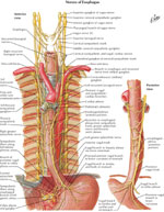
Figure 4
Parasympathetic and sympathetic innervation of the esophagus
Full size figure and legend (197K)
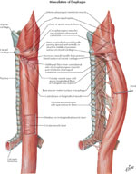
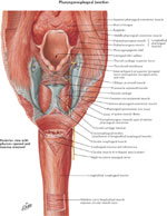
Figure 7
Upper esophageal sphincter and upper esophageal musculature
Full size figure and legend (151K)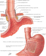
Figure 8
Gastroesophageal mucosal junction and muscular arrangement at the lower esophagus
Full size figure and legend (140K)
Figure 9
Diaphragmatic crura and esophageal opening viewed from below (a) and as viewed from above (b).
Full size figure and legend (114K)