
Figures, tables and video
From the following article
Surgical intervention and treatment of oral, pharyngeal motor disorders
Eugene A. Chu and James H. Kelly
GI Motility online (2006)
doi:10.1038/gimo51
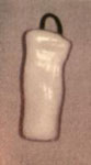
Figure 1
Weisberger and Huebsch endolaryngeal stent (Montgomery's stent) for treatment of chronic aspiration.
Full size figure and legend (36K)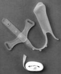
Figure 2
Eliachar endolaryngeal stent for treatment of chronic aspiration while allowing phonation.
Full size figure and legend (32K)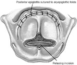
Figure 3
Supraglottic closure with epiglottic flap incorporating relaxing incision of epiglottis.
Full size figure and legend (56K)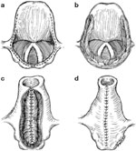
Figure 4
Biller's tube supraglottic laryngoplasty for supraglottic closure.
Full size figure and legend (76K)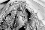
Figure 5
Intraoperative photograph of tubed supraglottic laryngoplasty.
Full size figure and legend (59K)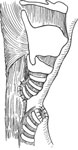
Figure 6
Tracheoesophageal diversion for subglottic closure and complete separation of the alimentary and respiratory passages.
Full size figure and legend (48K)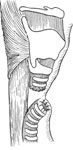
Figure 7
Laryngotracheal separation obviating the need for esophageal anastomosis by closure of the proximal tracheal stump.
Full size figure and legend (48K)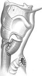
Figure 8
Modified tracheoesophageal diversion allowing esophageal anastomosis despite high tracheostomy.
Full size figure and legend (42K)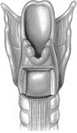
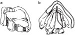
Figure 10
Partial cricoid resection modification of posterior cricoid resection.
Full size figure and legend (44K)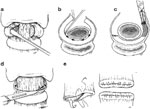
Figure 11
Subperichondrial cricoidectomy for definitive separation of the upper alimentary and respiratory passages.
Full size figure and legend (67K)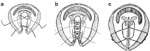
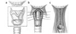
Figure 13
Narrow-field laryngectomy for definitive separation of the upper alimentary and respiratory passages.
Full size figure and legend (22K)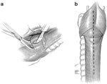
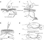
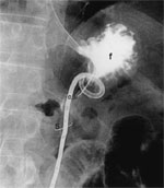
Figure 17
Radiograph of a Cope loop gastrostomy tube in position within the stomach.
Full size figure and legend (17K)
Table 1
Conditions associated with oral or pharyngeal motor dysfunction and dysphagia
Full size table and legend