Figures, tables and video
From the following article
Radiographic evaluation of motility of mouth and pharynx
Bronwyn Jones
GI Motility online (2006)
doi:10.1038/gimo25
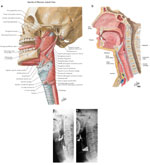
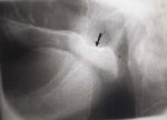
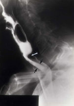
Figure 1
Lateral sagittal views of the pharynx comparing line drawings with contrast radiographs at rest and during phonation.
Full size figure and legend (235K)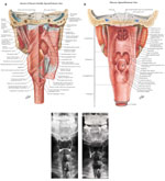
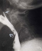
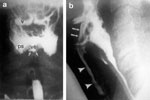
Figure 2
Comparisons between line drawings and radiographs in the coronal (frontal) plane.
Full size figure and legend (271K)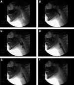
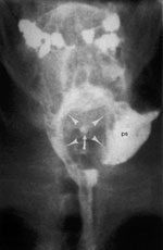
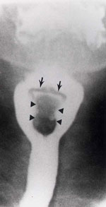
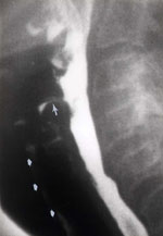
Figure 4
Selected stop-frame prints from a cinepharyngogram demonstrate several stages of a normal swallow.
Full size figure and legend (62K)