
Figure 2 - Comparisons between line drawings and radiographs in the coronal (frontal) plane.
From the following article
Radiographic evaluation of motility of mouth and pharynx
Bronwyn Jones
GI Motility online (2006)
doi:10.1038/gimo25
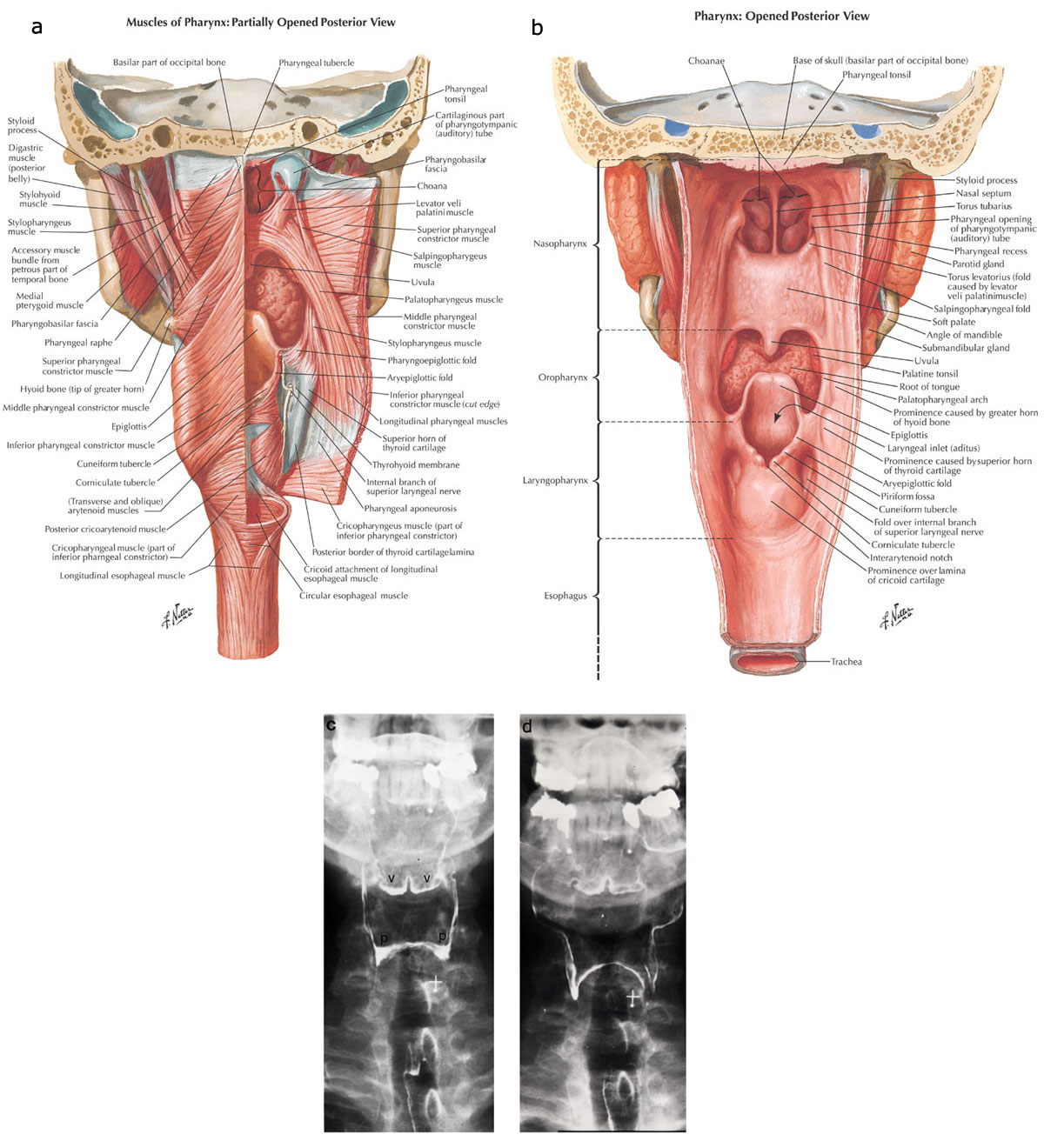
a: The constrictor and lateral suspensory muscles of the pharynx viewed from the posterior aspect, showing: the pharyngeal raphe, and the superior, middle and inferior pharyngeal constrictor muscles, the thyropharyngeus muscle and the cricopharyngeus muscle. Note again that the constrictor muscles overlap, the inferior being the more external and the superior the more internal. Note also that the fibers of the cricopharyngeus or horizontal fibers of the inferior constrictor muscle merge with the fascicles of the proximal circular muscle of the esophagus. b: The structures in the anterior wall of the pharynx as viewed from the posterior aspect. Note the uvula, epiglottis, piriform sinus and tongue. The mucosa remains intact. This drawing shows the contours of the valleculae and piriform sinuses and demonstrates the relationship of the valleculae to the base of the tongue and epiglottis. (a, b: Source: Netter images, with permission from Elsevier Science.) c: The pharynx has now been coated with high-density barium outlining the valleculae (v) and piriform sinuses (p). In this patient the epiglottis is not clearly visualized on this view at rest. d: The patient demonstrates "blowing up a balloon." The pharynx is now expanded especially in the region of the proximal portions of the piriform sinus. There is more symmetric distention proximally than distally because the superior portion lacks the support of the thyroid cartilage. (c,d: Source: Jones B, Donner MW. Examination of the patient with dysphagia. Radiology 1988;167:319–326, with permission from the Radiological Society of North America.)
Powerpoint slides for teaching
If the slide opens in your browser, Select "File > Save as" to save it.
Download Power Point slide (1,491K)