Figures, tables and video
From the following article
Oral, pharyngeal and upper esophageal sphincter motility disorders
Benson T. Massey and Reza Shaker
GI Motility online (2006)
doi:10.1038/gimo19

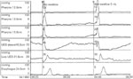
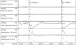
Figure 3
Manometric example of upper esophageal sphincter dysfunction.
Full size figure and legend (77K)