
Figures, tables and video
From the following article
Esophageal mucosal defense mechanisms
Roy C. Orlando
GI Motility online (2006)
doi:10.1038/gimo15
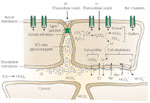
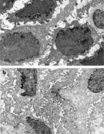
Figure 4
Diffusion of refluxed gastric acid (H+) into the intercellular space.
Full size figure and legend (62K)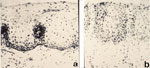
Figure 5
a: Normal esophageal suction biopsy from a healthy subject without esophagitis.
Full size figure and legend (44K)
Table 1
Factors contributing to esophageal mucosal defense against luminal acid
Full size table and legend