
Figures, tables and video
From the following article
Ivan M. Lang
GI Motility online (2006)
doi:10.1038/gimo12
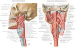
Figure 1
Anatomy of the closing and some opening muscles of the upper esophageal sphincter (UES).
Full size figure and legend (286K)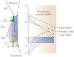
Figure 2
Relationship of upper esophageal high-pressure zone (UEHPZ) to the pharyngoesophageal muscles.
Full size figure and legend (37K)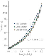
Figure 3
Strain-energy relationship of the cricopharyngeus (CP) muscle.
Full size figure and legend (33K)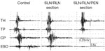
Figure 4
Effect of transection of various motor nerves on the motor responses of the UES closing muscles during swallowing.
Full size figure and legend (37K)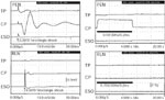
Figure 5
Effect of electrical nerve stimulation on electrical response and tension of UES closing muscles.
Full size figure and legend (85K)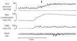

Figure 7
Relationship of CP and TP EMG activities to UES pressure during rest and excitation.
Full size figure and legend (67K)
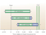
Figure 9
Temporal relationship of UES pressure, opening and CP EMG during 4-mL barium swallow relative to hyoid movement.
Full size figure and legend (34K)
Figure 10
Temporal relationship among UES pressure, trans-UES flow, and movement of the hyoid and larynx during 5-mL barium swallow.
Full size figure and legend (32K)
Figure 11
Movement of the hyoid bone during belching (a) and swallowing (b).
Full size figure and legend (27K)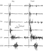
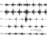
Figure 13
Responses of superior and inferior hyoid muscles during swallowing.
Full size figure and legend (45K)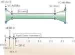
Figure 14
Temporal relationship among function of the glottis, UES, hyoid bone, and esophageal and stomach pressures during belching induced by rapid injection of 40 mL of air into the esophagus.
Full size figure and legend (16K)
Figure 15
Electromyography (EMG) responses of the opening and closing muscles of the UES during belching and swallowing activated by injection of 100 mL of air into the stomach of a chronically instrumented dog.
Full size figure and legend (42K)

Figure 17
Role of UES closure and opening muscles during the three phases of vomiting.
Full size figure and legend (56K)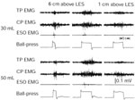
Figure 18
Effect of esophago-UES contractile reflex on the UES closure muscles.
Full size figure and legend (43K)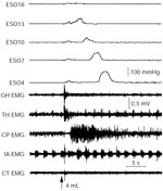
Figure 19
Role of UES closure muscles in esophago-UES relaxation reflex.
Full size figure and legend (61K)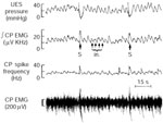

Table 1
Motor and sensory innervation of the muscles that open the upper esophageal sphincter (UES)
Full size table and legend