
Key Points
-
Provides an overview of the indications, strengths and limitations of the copy denture technique and outlines the key clinical and laboratory stages involved.
-
Highlights a range of different management strategies for patients with gag reflexes, for example, behaviour modification, distraction and systematic desensitisation.
-
Provides clinical advice on the management of anatomical challenges such as prominent tori and microstomia.
Abstract
The ability to provide high quality complete dentures remains an important skill for general dental practitioners. The prevalence of edentulism is increasingly concentrated within an older patient cohort and general dental practitioners may face challenges associated with providing care for these patients. This two-part series explores various aspects of complete denture provision and is designed to act as a refresher on the management of edentulous patients. This second part focuses on the copy denture technique as well as discussing strategies for assessing and managing gag reflexes, prominent palatal and lingual tori and microstomia.
Introduction
The first article in this two-part series outlined a variety of UK trends that will influence complete denture provision in the future. These include the concentration of edentulism within the oldest age groups and the challenges associated with transitioning to complete dentures later in life.1 In addition, negative societal perceptions of removable prostheses may increase patient expectations,2 whereas declining levels of undergraduate complete denture teaching may result in reduced confidence when managing edentulous patients in general dental practice.3,4,5 The first part also provided a refresher on the assessment and management of unstable lower dentures and fibrous replacement ridges. This second article will provide an update on the management of other conditions that may be encountered by a general dental practitioner including habituation, gagging, tori and microstomia.
Copy dentures
Introduction
Adapting to new dentures can be challenging for patients especially for those with long serving dentures.6 It has been suggested that older patients may experience difficulties in adapting to new dentures due to a reduced capacity for learning new muscle activity patterns.7 Anecdotal evidence suggests that utilising 'copy' or 'replica' techniques may help older patients adapt more easily to new dentures. These techniques allow favourable features from an existing denture to be copied (for example, the shape of the polished surfaces) while also allowing for alterations to less favourable features (for example, worn occlusal surfaces).8 There is, however, little evidence to suggest that patients will adapt to copy dentures any more successfully than to correctly constructed conventional dentures.9 Patient satisfaction with new complete dentures has a strong relationship with the quality of new dentures and also the residual mandibular alveolar ridge.10 Therefore, clinicians should focus on optimising denture design whether conventional or copy techniques are used.
Management
The decision to proceed with conventional dentures or copy dentures requires careful consideration of the patient's denture history and appraisal of the previous set of dentures.9 Copy denture techniques allow favourable features to be replicated while allowing minor improvements.8 Consequently, copy dentures are appropriate for situations where reduced adaptive capacity is presumed, only relatively minor alterations to the dentures are required, or when the appearance of the denture is to be replicated.6,8 It is likely that conventional processes will be more appropriate to dentures with significant deficiencies warranting greater changes to avoid the same errors being copied to the new set.8
Copy denture techniques typically involve fewer stages and less clinical time than conventional processes.9 Various authors have described modifications to the copy denture technique in the literature.8,9 One key variation relates to obtaining impressions before or after recording the occlusion. Clark et al. suggest that if the occlusion is recorded first, then the position of the replica denture bases will alter when the impression material is placed in them and would consequently be prone to occlusal errors.9 The following modifications to the copy denture technique are proposed:
-
First clinical stage: moulds of the original dentures are obtained in silicone putty (Fig. 1).9 If the dentures are underextended then the peripheral extension can be modified with a suitable material (for example, greenstick compound [ISO Functional Sticks, GC]) prior to taking the silicone mould6
Figure 1: Copy denture technique. 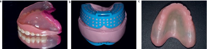
A decision was made to replicate the favourable features from the previous denture using the copy denture technique. a) The denture was initially border moulded using a thermoplastic material to improve the palatal extension. b) A silicone putty mould was obtained. c) Temporary rigid base with wax teeth was created. The wax teeth were subsequently adjusted to ensure correct occlusal vertical dimension, occlusal plane, centreline and bucco-lingual tooth positions. A closed mouth light bodied wash impression was then taken and a retruded jaw record was obtained
-
First laboratory stage: temporary rigid bases are produced in light cured or self-cured acrylic with wax teeth
-
Second clinical stage: the wax is adjusted to ensure correct occlusal vertical dimension, occlusal plane, centreline and bucco-lingual tooth positions.9 A closed mouth light bodied wash impression is taken with both maxillary and mandibular bases in situ. The retruded jaw record is obtained and a facebow record is made if appropriate
-
Second laboratory stage: the impressions are cast, articulated and a trial set up is constructed
-
Third clinical stage: the trial dentures are inserted, evaluated and modified as required. The position of the new post dam is scribed on the master model
-
Third laboratory stage: any final adjustments are undertaken and the dentures are processed. The dentures are removed from the flask, checked on the articulator for processing errors and polished
-
Fourth clinical stage: the dentures are checked intra-orally. A check record procedure may be undertaken as required.

Summary
Successful use of the copy denture technique relies on clinicians correctly diagnosing favourable and unfavourable features of previous dentures. This technique provides benefits through fewer clinical stages. Anecdotal evidence suggests that copying features from old dentures may help older patients adapt more easily to new dentures. However, the technique is inappropriate if there are any major inadequacies with previous dentures. Clinicians should focus on optimising support, retention and stability regardless of whether conventional or copy techniques are undertaken.
Gagging
Introduction
Gagging is a common problem faced by clinicians and patients. It can impede the effective delivery of dental procedures and may lead to non-attendance or avoidance of preventative measures.11,12,13,14 It is therefore important to understand the aetiology of this phenomenon to enable the provision of high quality dental care. Gagging is a broad term used to describe a 'normal' defence mechanism that prevents foreign bodies from entering the trachea, pharynx, or larynx.11 Sakamoto et al. describe the response via two additive mechanisms.15 Firstly, a physiological response related to afferent nerve impulses and secondly, an emotional response thought to result in stimulation of the gagging centre via sympathetic nervous activation. Both mechanisms are important as different factors may play differing roles in individual patients. The gag reflex correlates strongly with high levels of anxiety trait.11
Classification
In the context of complete denture provision, a gag reflex is generally assessed by determining whether the entire maxillary denture bearing area can be palpated with a finger without triggering the gag reflex. There are also a number of classification systems which exist for assessing gagging from the perspective of providing general dental treatment. The Classification of Gagging Problem (CGP) index is summarised in Table 1.15,16 This provides a relatively simple and user-friendly assessment, however, it is subject to bias due to the lack of explicit definition of clinical parameters. For example clinicians may position a dental mirror differently when assessing the same teeth. Fiske and Dickinson published a Gagging Severity Index in 2001 which is at risk of similar sources of bias.17 Furthermore, various tools exist for assessing the severity of a gag reflex from the patient's self-reported perspective, for example, the Gagging Problem Assessment questionnaire (GPA).18,19 Although not commonly used in normal clinical practice, these indices may be useful to help assess or monitor the severity of a gag reflex.
Management
Patients may be divided into three broad management categories: a) those that are able to tolerate impressions with distraction; b) those that are unable to tolerate impressions and need pharmacological aids (for example, sedation) to enable impressions to be undertaken; and c) those that are unable to tolerate the dentures and may need modifications to the prostheses. The following sections provide an overview of some useful techniques that may be implemented when managing patients with a gag reflex.
Behaviour modification
The vast majority of patients with a gag reflex will contribute cognitively to the gagging response via the emotional response pathway.20 Consequently, it has been suggested that all disruptive gagging should be presented to the patient as a behavioural response which is amenable to modification.20 Behaviour modification is reported to be the most successful long-term management for gagging.21 This approach is outlined in Figure 2.

Pathway for assessing gag reflex in the context of the proposed dental treatment
Impression techniques and distraction
It is important to build a good rapport with the patient and adopt a positive and sympathetic approach to help build the patient's confidence with dental procedures. Patients should be reassured that gagging is commonly encountered by dental professionals and can often be managed by simple techniques. Prior to impression taking, both stock and special trays should be assessed for overextension and trimmed if overextended. An impression material with a thicker consistency and shorter set time may be beneficial for patients with a gag reflex. For primary impressions, a thermoplastic impression material (for example, Impression Compound, Kerr) may be useful as the tray can be readily removed during a gagging episode and the material can be repeatedly warmed, soften and reseated until the desired impression is obtained. Alternatively, if using alginate, mixing the material with warm water will shorten the set time and a stiffer mix is likely to be more tolerable to the patient.
Impression materials should be carefully handled, with care taken to avoid overloading the impression trays. The loaded tray should be first seated at the posterior aspect before seating the tray anteriorly to ensure that excess material is not expressed at the posterior aspect of the tray. Patients should be reassured throughout the impression procedure and encouraged to perform basic breathing, relaxation or distraction exercises. Patients should be encouraged to tilt their head forwards and breathe through their nose if they find this is more comfortable. A saliva ejector can be utilised to aspirate any excess saliva. Careful handling of impression material and constant patient reassurance or distraction may be all that is necessary to obtain a satisfactory impression.
Relaxation, hypnotherapy and sedation
Relaxation techniques for managing anxiety and aberrant behaviours (including gagging) have been researched in many fields. This can be useful in managing unhelpful thought processes and help the patient gain control over an unpleasant event such as a dental impression. It has also been suggested that adjuncts such as hypnotherapy and acupuncture can be useful in the management of a gag reflex.22,23 Clinical experience suggests that this is rarely necessary if other techniques are employed correctly. Some of these techniques only temporarily increase compliance and may therefore have limited utility in denture provision (for example, acupuncture). Furthermore, sedation techniques may be utilised to aid impression taking. However, techniques which impair patient awareness are only suitable with appropriate training, planning and consent.22,23,24
Systematic desensitisation
Where patients are unable to tolerate a prostheses, systematic desensitisation techniques can be useful to slowly increase a patient's exposure to a stimulus that would normally trigger gagging.25 Many different desensitisation techniques have been described.20,26,27 Contemporary techniques involve the patient holding a toothbrush or empty stock tray in their mouth and progressing with location targets (for example, inserting further posteriorly) or time targets (for example, retaining for longer). One of the most commonly utilised techniques involves acrylic training plates which comprise a well retained and stable denture plate base that the patient wears for increasing time periods (Fig. 3).25 The base plate should ideally be constructed of a rigid material such as clear heat cure acrylic to ensure optimal fit. It often has multiple post dams to allow for modification and can be supported with an anterior rim to facilitate placement and removal thus ensuring patient control.25 On occasion, denture teeth can be added to the training plate to help with motivation.

a) An example of a clear acrylic training plate with multiple post dams to allow for modification. b) Once the optimal extension of the training plate had been determined, progression with complete denture construction was possible. The denture base extension matched the training plate
With systematic desensitisation, patient communication and planning are key to ensure adequate progress is achieved and the patient does not become disheartened or frustrated with either an overly lengthy process or one where the patient is required to adapt to large changes too quickly. Both the clinician's and patient's expectations must be aligned with the patient's ability to adapt. A controlled stepwise approach with individual targets is usually recommended although some authors recommend more regimented protocols.11 Many authors suggest patients wear their desensitising training plates when they are otherwise occupied or distracted.25 Once the optimal extension of the training plate is determined, progression with complete denture construction is often possible and the denture base extension can be designed to match the existing training plate extension.
Horseshoe-shaped maxillary dentures
Patients may request the construction of a palateless or horseshoe-shaped maxillary denture with the expectation that this would help with their gag reflex. Unfortunately, these dentures often have compromised stability and retention as a result of the reduced palatal extension. This approach is therefore more likely to stimulate a gag reflex and will often result in a compromised outcome with patient dissatisfaction.20,28,29 Some authors therefore recommend the construction of a horseshoe-shaped maxillary denture with a severely reduced palatal extension only when other treatment modalities have failed.30
Implant-supported rehabilitation
In severe cases it may be appropriate for the patient to receive implant-supported rehabilitation. It is important to note that these cases may still require some attenuation of a gag reflex to ensure they are able to both undergo treatment procedures associated with implant placement and restoration, in addition to tolerating the final prosthesis. Nonetheless, an implant-supported rehabilitation may be beneficial in patients who are unable to progress further with the management strategies described above but would be able to tolerate a palateless overdenture.
Summary
Gagging is a common problem that may be influenced by physiological or psychological factors. Management strategies should be tailored to the individual and may comprise a combination of behaviour modification, distraction and systematic desensitisation. These should be clear, managed collaboratively with the patient, and supported by a good patient-clinician rapport.
Tori
Introduction
Tori are bony protrusions that constitute normal anatomy. They can affect the palate (where they often present in the midline), or the mandible (where they often present symmetrically affecting the lingual alveolus). They are diagnosed based on clinical examination and are often consistent with healthy soft tissues with single or multiple bony protuberances. History will confirm an unchanging appearance over time.
Classification
There is a lack of research to provide an evidence-based classification of tori. Management is usually based on clinical characteristics identified during assessment. It should be noted that radiographic assessment of tori is usually not necessary as the information is unlikely to influence diagnosis or management unless surgical intervention is being considered. In such an eventuality it is important to conform to principles of radiographic exposures for medical investigations.
Location
Palatal tori typically present as a solitary mass in the midline of the palate and are most often symmetrical, although slight variation on this may occur. Larger palatal tori may present with a central fissure or a bilobed appearance. Lingual tori typically present as two or more masses in a symmetrical pattern in the canine/premolar region. Lingual tori can be sufficiently prominent that their incorporation into the denture base design is ill-advised due to the impact on comfort and invasion of the tongue space.
Condition of overlying tissues
Soft tissues overlying tori are often healthy but may present with areas of displaceable mucosa overlying bony prominences or undercuts. It is also important to examine for signs of inflammation, ulceration or overgrowth which may be attributable to denture trauma or stomatitis.31,32 Such problems should be effectively managed before undertaking master impressions.
Contour
The contour of tori should be considered to determine the likely impact on the path of insertion of a prosthesis and the need to block out undercuts. Undercuts may present on the posterior edges of palatal tori and inferior edges of lingual tori. The potential impact of the tori in relation to anticipated soft tissue undercuts and path of insertion may be easier to evaluate on a primary cast.
Management
Relief of tori
Historically there has been debate as to whether tori should be relieved during the denture manufacturing process. Previous clinical guidance suggests that relief of tori is not necessary.31 However, relief can be provided where there is concern that the morphology may result in trauma to the overlying tissues during denture wear for example, in cases with significant undercuts or thin overlying mucosa. Relief is typically performed by applying foil (usually 0.5 mm thickness) over areas of interest on the working cast. This is then carefully adapted to the soft tissue contour and burnished. Relief is often only provided over areas where it is deemed necessary.
Denture border extension
Minor palatal tori can often be incorporated into the design of a denture by extending the major connector over the torus. Large palatal tori may significantly reduce the available surface area for retention, support and bracing of a maxillary denture and may be impractical to cover. There is a lack of research reporting on the management of such cases. While not conventional practice, some authors have utilised ring connectors with a window in the palate to accommodate a large palatal torus.33 This type of design is likely to compromise maintenance of a border seal and impair retention.
The location of a torus may compromise the posterior border seal by significantly reducing the anteroposterior dimension of the major connector. High impact acrylic or a metal denture base should be considered where the mechanical integrity of the major connector is compromised – this should be balanced with the relative weight of each material and the anticipated retention of the prosthesis. Figure 4 highlights the role of these factors in effective treatment planning.

a & b) A patient presented with a large palatal torus extending close to the anticipated post-dam region. c) The previous horse-shoe shaped maxillary denture provided unsatisfactory retention and bracing. d) A new full coverage complete denture was designed to optimise retention and bracing. Care was taken to ensure acrylic was of sufficient thickness in the post-dam region and not too bulky over the torus. High impact acrylic offered rigidity and strength
Lingual tori may be sufficiently prominent that their incorporation into the denture base design is ill-advised due to the impact on both comfort and invasion of the tongue space. In such cases, they are often avoided in the design of the major connector. It may be necessary to utilise a denture base material with better mechanical properties in narrow section (for example, cobalt-chrome). Surgical intervention may be rarely considered, although the risks of this procedure must be weighed against the benefits to any rehabilitation and subsequent quality of life in the long-term.
Surgical removal
The surgical removal of tori is rarely necessary and there is a lack of consensus or high-quality evidence to guide planning. If surgical removal is being considered, then referral to specialist services should be made for further assessment. This would involve an initial prosthodontic assessment, followed by surgical and radiographic assessment if surgical intervention is considered.
Summary
Tori should be managed on a case-by-case basis following assessment of the location, relationship to denture borders, contour and the condition of the overlying tissues. Minor tori can often be incorporated into the denture design and covered with the denture base whereas larger palatal or lingual tori may need more significant modifications. It is important to remember that a staged approach is often necessary which will involve the provision of dentures with optimised coverage in the first instance. Should this prove unsuccessful, alternative approaches may be necessary such as provision of dentures with compromised tissue coverage or in rare cases surgical removal of tori.
Microstomia
Introduction
Microstomia is defined as an abnormally small oral orifice34 and can be associated with a variety of acquired or congenital conditions.35 Microstomia can be a consequence of facial burns, the management of head and neck cancer, or scarring following surgery or trauma.35,36,37 It can also present as a clinical manifestation of systemic connective tissue diseases (such as scleroderma) or congenital syndromes.37,38 Restricted oral access can complicate oral hygiene, provision of dental treatment and prosthetic rehabilitation.36,38 It may also result in functional difficulties such as speech impairment, distortion of facial expression and difficulties with mastication and deglution.36,38
Classification
Some authors measure the intrinsic vertical mouth opening as an indication of the severity of microstomia. It has been suggested that the average intrinsic vertical mouth opening measures 40–50 mm, a reduced opening of 25–35 mm is “functional” and an opening of 10–24 mm is “severely limiting”.36 Additionally, an index of oral access has been proposed to aid clinicians in diagnosing, recording, treatment planning and monitoring the severity of microstomia.35 This grades the severity of access for restorative dental treatment depending on whether the clinician can access all areas of the dentition or if modifications are necessary to facilitate impressions and prosthesis design.35
Management
General management
General management strategies for improving the oral aperture will vary depending upon the aetiological factors and severity of symptoms. Conservative management strategies include scar massage, daily stretching exercises and use of oral stretching devices.36,39 Surgical approaches (for example, commisuroplasties) may also be advocated in severe cases which are refractory or not amenable to conservative management.36
Prosthetic management
Provision of removable prostheses can be challenging for patients with microstomia. In addition, patients may experience other factors associated with the aetiology which would further limit their ability to tolerate dentures. For example, patients with systemic sclerosis may suffer from xerostomia, mucosal ulceration, and reduced manual dexterity.38
In patients with mild – moderate microstomia, relatively simple modifications may be required to facilitate denture construction (Fig. 5). Impressions may be aided by reducing the height of the impression trays, using a rotational path of insertion, applying petroleum jelly to the commissures and asking the patient to half close their mouth.37 At the wax try-in stage, the positioning of the denture teeth should be carefully appraised. A compromise may need to be made between optimal aesthetics/lip support and the likelihood of denture displacement by the fibrotic tissues. The size of the denture teeth and occlusal table should be reduced appropriately.40

a & b) 56-year-old patient with systemic sclerosis. Complete denture construction had been unsuccessful in general practice due to her reduced oral aperture. c) Significant modification was required to the stock trays to allow insertion. d & e) Successful rehabilitation with complete dentures was possible, however, some compromises were necessary eg, accepting a class III incisal relationship and reduced lip support. e) The final denture which was appropriately extended to avoid impinging on the fibrous tissues
In more severe cases, more significant modification may be required. Where a patient has previously successful complete dentures, a copy denture technique may be useful.37 Sectional impression trays have been advocated in the literature for both primary and secondary impressions.41 These tend to record the ridge in two parts and can be disassembled before relocating them outside the mouth.37,41
The use of sectional or collapsible denture materials have been reported to aid denture insertion. These reports have used a range of mechanisms to connect the different parts of the denture including clasps, cobalt-chrome hinges, swing-lock attachments, stud attachments, rods and magnets.35,42,43 Construction of these dentures can involve more complex clinical and laboratory stages compared to conventional dentures and would require increased manual dexterity for insertion. In addition, some case reports have utilised flexible denture materials either in isolation44 or in conjunction with rigid superstructures to rehabilitate patients with microstomia.35 The literature supporting the use of flexible denture base materials in such cases is limited in relation to longevity.45,46
Summary
The effective management of patients with microstomia should be based on careful assessment of the degree of oral access and anticipation of other potential challenges associated with its cause. Provision of removable prostheses for patients with mild microstomia may be successfully managed in general dental practice with relatively simple adjustments to impression technique. More severe cases may need referral for specialist input due to more complex impression and prosthesis manufacturing procedures. In extreme cases, prosthetic rehabilitation may not be possible.35
Conclusion
Complete dentures have been the traditional standard of care for edentulous patients for many years and for many patients, this has allowed them to function in society more easily than without any prosthesis.47 It is well recognised that many people struggle with complete dentures and throughout this two-part series we have discussed a range of different factors that may impact on a patient's ability to manage removable prostheses.
Consensus suggests that the restoration of the edentulous mandible with a conventional denture is no longer the most appropriate first choice prosthodontic treatment.47 A substantial body of evidence demonstrates greater patient satisfaction and quality of life with implant-supported dentures than for conventional dentures.48 Despite this, uptake of implant technology for complete denture wearers has been slow.48 Some patients may not be suitable for implant-supported rehabilitation while others may find this option is prohibitively expensive.
The importance of understanding and applying the principles of complete denture construction remains vital in providing an appropriate quality of care to patients. The general dental practitioner should be able to assess different anatomical, physiological, pathological or psychological factors that may impact on complete denture construction. This will enable the development of an appropriate management strategy and facilitate informed consent. Many of these principles of complete denture construction will also be important with removable implant-retained overdentures.
References
Information Centre for Health and Social Care, Office for National Statistics. Social Survey Division. (2012). Adult Dental Health Survey, 2009. [data collection]. Second Edition. UK Data Service. SN: 6884.
Bradnock G, White D A, Nuttall N M, Morris A J, Treasure E T, Pine C M . Dental attitudes and behaviours in 1998 and implications for the future. Br Dent J 2001; 190: 228–232.
Clark R K, Radford D R, Juszczyk A S . Current trends in complete denture teaching in British dental schools. Br Dent J 2010; 208: E10; discussion 214–215.
Wieder M, Faigenblum M, Eder A, Louca C . An investigation of complete denture teaching in the UK: part 1. A survey of undergraduate teaching. Br Dent J 2013; 215: 177–181.
Wieder M, Faigenblum M, Eder A, Louca C . An investigation of complete denture teaching in the UK: part 2. The DF1 experience. Br Dent J 2013; 215: 229–236.
Beddis H P, Morrow L A . Technique tips-greenstick modification of dentures Prior to the replica technique: 'how we do it'. Dent Update 2013; 40: 688.
Brill N, Tryde G, Schübeler S . The role of learning in denture retention. J Prosthet Dent 1960; 10: 468–475.
McCord J F, Hannah V E, Cameron D, Watson D, Donaldson A C . An update on the replica denture technique. Dent Update 2010; 37: 230–232, 235.
Clark R K, Radford D R, Fenlon M R . The future of teaching of complete denture construction to undergraduates in the UK: is a replacement denture technique the answer? Br Dent J 2004; 196: 571–575.
Fenlon M R, Sherriff M . An investigation of factors influencing patients' satisfaction with new complete dentures using structural equation modelling. J Dent 2008; 36: 427–434.
Bassi G S, Humphris G M, Longman L P . The aetiology and management of gagging: a review of the literature. J Prosthet Dent 2004; 91: 459–467.
Dickinson C M, Fiske J . A review of gagging problems in dentistry: I. Aetiology and classification. Dent Update 2005; 32: 26–28, 31–32.
Dickinson C M, Fiske J . A review of gagging problems in dentistry: 2. Clinical assessment and management. Dent Update 2005; 32: 74–76, 78–80.
Scarborough D, Bailey-Van Kuren M, Hughes M . Altering the gag reflex via a palm pressure point. J Am Dent Assoc 2008; 139: 1365–1372.
Sakamoto T, Fukuda K, Saita N et al. Autonomic nervous activity of patients with gagging problems during dental mirror insertion. Spec Care Dent 2016; 36: 80–84.
Saita N, Fukuda K, Koukita Y, Ichinohe T, Yamashita S . Relationship between gagging severity and its management in dentistry. J Oral Rehabil 2013; 40: 106–111.
Fiske J, Dickinson C . The role of acupuncture in controlling the gagging reflex using a review of ten cases. Br Dent J 2001; 190: 611–613.
Akarslan Z Z, Bicer A Z . Utility of the gagging problem assessment questionnaire in assessing patient sensitivity to dental treatments. J Oral Rehabil 2012; 39: 948–955.
Van Linden van den Heuvell G F, Ter Pelkwijk B J, Stegenga B . Development of the Gagging Problem Assessment: a pilot study. J Oral Rehabil 2008; 35: 196–202.
Ramsay D S, Weinstein P, Milgrom P, Getz T . Problematic gagging: principles of treatment. J Am Dent Assoc 1987; 114: 178–183.
Altamura L S, Chitwood P R . Covert reinforcement and self-control procedures in systematic desensitization of gagging behaviour. Psychol Rep 1974; 35: 563–566.
Barsby M J . The use of hypnosis in the management of 'gagging' and intolerance to dentures. Br Dent J 1994; 176: 97–102.
Robb N D, Crothers A J . Sedation in dentistry. Part 2: Management of the gagging patient. Dent Update 1996; 23: 182–186.
Noble S . The management of blood phobia and a hypersensitive gag reflex by hypnotherapy: a case report. Dent Update 2002; 29: 70–74.
Ali R, Altaie A, Morrow L . Prosthetic rehabilitation of the gagging patient using acrylic training plates. Dent Update 2015; 42: 52–54, 56–58.
Conny D J, Tedesco L A . The gagging problem in prosthodontic treatment. Part II: Patient management. J Prosthet Dent 1983; 49: 757–761.
Singer I L . The marble technique: a method for treating the “hopeless gagger” for complete dentures. J Prosthet Dent 1973; 29: 146–150.
Akeel R, Assery M, al-Dalgan S . The effectiveness of palate-less versus complete palatal coverage dentures (a pilot study). Eur J Prosthodont Restor Dent 2000; 8: 63–66.
Zach GA . Gag control. Gen Dent 1989; 37: 508–509.
Farmer J B, Connelly M E . Palateless dentures: help for the gagging patient. J Prosthet Dent 1984; 52: 691–694.
McCord J F, Grant A A . A Clinical Guide to Complete Denture Prosthodontics. British Dental Association, 2000.
McCord J F, Grant A A . Identification of complete denture problems: a summary. Br Dent J 2000; 189: 128–134.
Rajeev V, Arunachalam R . Innovative replication and recuperation of complex torus palatinus: A prosthodontic case report. World J Dent 2016; 7: 208–212.
The Glossary of Prosthodontic Terms: Ninth Edition. J Prosthet Dent 2017; 117: e1–e105.
King E, Owens J . Flexible and Sectional Complete Dentures with Magnetic Retention for a Patient with Microstomia-A Case Report. Dent Update 2016; 43: 212–213.
Zweifel C J, Guggenheim M, Jandali A R, Altintas M A, Kunzi W, Giovanoli P . Management of microstomia in adult burn patients revisited. J Plast Reconstr Aesthet Surg 2010; 63: e351–e357.
Garnett M J, Nohl F S, Barclay S C . Management of patients with reduced oral aperture and mandibular hypomobility (trismus) and implications for operative dentistry. Br Dent J 2008; 204: 125–131.
Veale B J, Jablonski R Y, Frech T M, Pauling J D . Orofacial manifestations of systemic sclerosis. Br Dent J 2016; 221: 305–310.
Dougherty M E, Warden G D . A thirty-year review of oral appliances used to manage microstomia, 1972 to 2002. J Burn Care Rehabil 2003; 24: 418–431.
Alantar A, Cabane J, Hachulla E et al. Recommendations for the care of oral involvement in patients with systemic sclerosis. Arthritis Care Res (Hoboken) 2011; 63: 1126–1133.
Hegde C, Prasad K, Prasad A, Hegde R . Impression tray designs and techniques for complete dentures in cases of microstomia-a review. J Prosthodont Res 2012; 56: 142–146.
Colvenkar S S . Sectional impression tray and sectional denture for a microstomia patient. J Prosthodont 2010; 19: 161–165.
Watanabe I, Tanaka Y, Ohkubo C, Miller A W . Application of cast magnetic attachments to sectional complete dentures for a patient with microstomia: a clinical report. J Prosthet Dent 2002; 88: 573–577.
Egan J G, Swindells S A . A novel prosthodontic alternative for patients who are edentulous and have microstomia: a case report. Spec Care Dentist 2012; 32: 160–164.
Polychronakis N C, Polyzois G L, Lagouvardos P E, Papadopoulos T D . Effects of cleansing methods on 3-D surface roughness, gloss and colour of a polyamide denture base material. Acta Odontol Scand 2015; 73: 353–363.
Vojdani M, Giti R . Polyamide as a Denture Base Material: A Literature Review. J Dent 2015; 16: 1–9.
Feine J S, Carlsson G E, Awad M A et al. The McGill consensus statement on overdentures. Mandibular two-implant overdentures as first choice standard of care for edentulous patients. Gerodontology 2002; 19: 3–4.
Thomason J M, Feine J, Exley C et al. Mandibular two implant-supported overdentures as the first choice standard of care for edentulous patients-the York Consensus Statement. Br Dent J 2009; 207: 185–186.
Author information
Authors and Affiliations
Corresponding author
Rights and permissions
About this article

Cite this article
Jablonski, R., Patel, J. & Morrow, L. Complete dentures: an update on clinical assessment and management: part 2. Br Dent J 225, 933–939 (2018). https://doi.org/10.1038/sj.bdj.2018.1023
Accepted:
Published:
Issue Date:
DOI: https://doi.org/10.1038/sj.bdj.2018.1023
This article is cited by
-
The impact of a quality improvement initiative to reduce denture loss in an acute hospital
British Dental Journal (2022)
-
Implant-supported overdentures: part 2
British Dental Journal (2021)
