Abstract
When nutrients run out, bacteria enter a dormant metabolic state. This low or undetectable metabolic activity helps bacteria to preserve their scant reserves for the future needs, yet it also diminishes their ability to scan the environment for new growth-promoting substrates. However, neighboring microbial growth is a reliable indicator of a favorable environment and can thus serve as a cue for exiting dormancy. Here we report that for Escherichia coli and Pseudomonas aeruginosa this cue is provided by the basic peptidoglycan unit (i.e. muropeptide). We show that several forms of muropeptides from a variety of bacterial species can stimulate growth resumption of dormant cells and the sugar – peptide bond is crucial for this activity. These results, together with previous research that identifies muropeptides as a germination signal for bacterial spores, and their detection by mammalian immune cells, show that muropeptides are a universal cue for bacterial growth.
Introduction
Free-living bacteria typically encounter large fluctuations in their environment such as the transition from nutrient abundance to scarcity, i.e. feast and famine cycle. When growth substrates are exhausted, bacteria initiate specific developmental programs that prepare them for a long period of dormancy. Many Gram-positive bacteria form spores that are very resilient to adverse conditions and can survive hundreds of years1. The morphological transition of Gram-negative bacteria into dormant cells is perhaps less drastic, but significant changes do occur.
Gram-negative bacteria undergo large changes in their gene expression pattern and metabolism when entering stationary phase2. These changes are largely governed by the alarmone (p)ppGpp that alters the transcription of many genes, including reducing growth-oriented and increasing survival-oriented gene expression3,4,5. Ribosome synthesis decreases and, later, inactive 100 S ribosomal particles form. DNA replication is also inhibited. At the end of these changes, cells enter a dormant state able to withstand long period without nutrients.
Much less is known about recovery from dormancy when nutrients become available again. It is now clear that cells display considerable phenotypic heterogeneity in the timing of recovery – in clonal population some cells start growing rather quickly while others stay dormant for longer and initiate growth only later6,7. The timing of growth resumption has been suggested to rely on a stochastic process, but some reports also describe different states of dormancy (shallow and deep dormancy) and suggest that growth resumption from shallow dormancy is quicker8,9. We have shown that the order of cells resuming growth under some conditions is determined by the order they enter stationary phase, indicating a long-term memory effect in E. coli10.
Persisters are antibiotic tolerant cells in a generally antibiotic sensitive bacterial population11. Consensus is now emerging that persisters are mostly non-dividing cells that survive antibiotics due to their inactivity12. They only recover from dormancy and start to grow after a long lag phase that exceeds the antibiotic treatment regime. Given that persisters are held responsible for several recurrent infections13,14 it is important to understand the mechanisms that govern the resumption of growth.
The speed of growth resumption can be influenced by the environment. E. coli recovery from stationary phase is quicker in rich media and leads to fewer antibiotic tolerant cells6. Slow recovery in the presence of a non-optimal carbon source can be accelerated by small amount of glucose that probably acts as a signal rather than a nutrient10. In Micrococcus luteus growing cells secrete an Rpf protein that can induce growth resumption of dormant cells15 and dormant Staphylococcus aureus cells can be resuscitated with the help of spent culture supernatant16. Bacillus spores are able to detect nutrients and other molecules through specific receptors and initiate germination in response17,18. Here we describe a growth resumption signal for E. coli consisting of muropeptides. These molecules are produced by actively growing cells and act to stimulate the growth resumption of dormant cells. Moreover, dormant cells from both E. coli and Pseudomonas aeruginosa resume growth in response to muropeptides from other species, thereby indicating their role as an interspecies signal. We describe the structural requirements for muropeptide activity and isolate mutants with altered sensitivity to muropeptides.
Results
Dividing cells secrete a factor that stimulates growth resumption
During our studies on growth resumption heterogeneity10 we speculated that growing cells produce a signal that stimulates the growth resumption of non-growing cells. To directly test this hypothesis we prepared a conditioned medium and tested its effect on cells that resume growth from stationary phase. Stationary phase cells were washed and resuspended in fresh medium. Half of the culture was immediately centrifuged and the supernatant was sterilized by filtration for use as a control medium. Prepared in this way, the control medium is almost like fresh medium, but also contains possible residual metabolites carried over by cells from stationary phase. The other half were grown until the middle exponential phase (OD (at 600 nm) about 0.5, while stationary phase OD is 1.0) and used for the preparation of conditioned medium. This conditioned medium should still have enough substrates to support growth and also contain factors secreted, if any, by growing cells.
We then took cells from stationary phase and compared their growth resumption in fresh, control, and conditioned media. At first we used our single-cell growth resumption assay10 where cells, carrying two plasmids encoding for fluorescent proteins GFP and Crimson, are grown into stationary phase with Crimson expression induced by arabinose (Fig. 1a). After that the arabinose is removed and a fresh carbon source is added together with IPTG to induce GFP. Cells that resume growth, begin active protein synthesis and initially become GFP-positive and later dilute their Crimson levels by cell division. GFP-positive cells clearly accumulate quicker in conditioned medium (Fig. 1b). The same effect is evident when comparing the optical density of different cultures – the lag phase is shorter in conditioned medium (Fig. 1c).
Conditioned medium stimulates growth resumption of stationary phase E. coli. (a) A scheme for single-cell growth resumption assay on flow cytometer. (b) Stationary phase cells were resuspended either in conditioned medium or control medium and GFP expression was induced. Growth resuming cells are GFP-positive and dividing cells have reduced Crimson content due to dilution by cell division. (c) Stationary phase cells were diluted into fresh medium, and fresh medium mixed with conditioned medium or control medium. Cells were grown in 96-well plate and OD was measured every 15 minutes. Cells exposed to conditioned medium resume growth earlier than cells in fresh medium. (d) Quantification of results in panel b. Δt is the time difference (in minutes) of reaching OD 0.06 (actual reading for 100 μl culture) between test medium and fresh medium. Dilution factor indicates how many times the tested medium was diluted in fresh medium. The average of three independent experiments are shown, error bars indicate standard error of the mean.
Compared to fresh medium, growth resumption is slightly inhibited in control medium. This is probably due to some inhibitory compounds carried over by stationary phase cells (see the preparation method above). At the same time the growth rate in exponential phase was the same for all cultures, thus indicating that only the growth resumption was affected. For quantification we compared the time it took for different cultures to reach OD 0.06 (measured OD value for 100 μl culture on 96-well plate at 600 nm, chosen to be approximately in the middle of the exponential phase) and calculated the difference between conditioned (or control) medium and the culture grown in fresh medium (Δt). The growth stimulatory effect of the conditioned medium was concentration dependent and still clearly detectable when diluted several times (Fig. 1d). These results demonstrate that conditioned medium has growth resumption promoting activity.
Cell wall derived muropeptides stimulate growth resumption
We tried to purify this activity from conditioned supernatant, but failed to get enough pure material for identification. However, during this process we learned that the molecule that facilitates growth has a relatively low molecular weight, is hydrophilic, and not strongly charged. Because cell wall derived muropeptides (MPs) fit this description and can induce spore germination in Bacillus18, we went on to test if MPs can also stimulate growth resumption in E. coli.
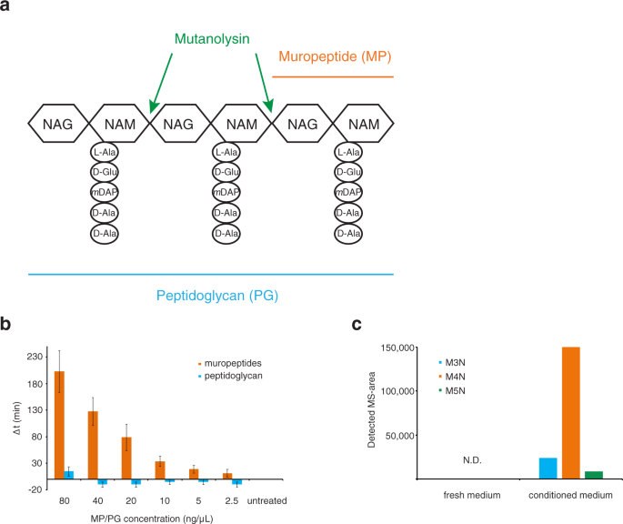
For this, we purified peptidoglycan (PG) from growing E. coli cells using an established method18 and digested it with mutanolysin to obtain individual MPs that consist of a disaccharide (N-acetyl-glucosamine linked to N-acetyl muramic acid) and a short peptide bound to muramic acid (Fig. 2a). When added to fresh medium, these solubilized MPs convey growth resumption stimulating activity whereas undigested PG does not (Fig. 2b).
Muropeptides stimulate growth resumption of E. coli. (a) Schematic representation of peptidoglycan and muropeptide. NAG – N-acetyl-glucosamine, NAM – N-acetyl-muramic acid. (b) Muropeptides (MPs) but not peptidoglycan (PG) can stimulate growth resumption of stationary phase cells in fresh medium. Different amounts of MP or PG were added to recovering cells and the Δt was calculated. The average and standard error of the mean of four independent experiments are shown. (c) Using mass-spectrometry, different MP variants can be detected from conditioned medium, but not from fresh medium. M4N – anhydromuro-tetrapeptide, M3N – anhydromuro-tripeptide, M5N – anhydromuro-pentapeptide, N.D. – not detected. The values shown are an average of two technical replicates.
In order to allow cell enlargement, peptidoglycan hydrolases cleave the PG sacculus during cell growth to generate PG fragments. The MPs released are usually recovered – both Gram-positive and Gram-negative bacteria have MP recycling systems that transport PG fragments back to their cytoplasm where they can be re-used19,20. However, some MPs escape the transport system and diffuse throughout the growth environment. We analyzed if there are MPs in our conditioned medium. For this, fresh and conditioned medium was concentrated and subjected to UPLC-MSe analysis. We detected anhydro disaccharide with tri-, tetra-, or pentapeptides in the conditioned medium, but not in fresh medium (Fig. 2c). This is in line with previous results21 and further supports our hypothesis that MPs act as a growth resumption signal in the conditioned medium.
It is necessary to observe that the effect of MPs is evident only when the lag phase is long enough. A longer stationary phase, good aeration during the stationary phase, and the use of “non-optimal” carbon sources in the growth resumption medium all prolong the lag phase and expose the effect of MPs. When stationary phase cells are resuspended in favorable media (LB or MOPS glucose), all cells resume growth quickly and the effect of MPs is not detectable on this background.
Bacterial PG is quite well conserved across different taxa and its basic structure is the same in most species22. It is thus reasonable to assume that E. coli growth resumption could also be stimulated by MPs from other species. One noticeable difference between the PG composition from Gram negatives and some Gram positives is the amino acid present at the third position of the peptide stem: Gram negatives, including E. coli, usually have meso-diaminopimelic acid (mDAP) in that position, while most Gram positives contain L-lysine (Lys)22. We prepared MPs from Gram positive bacteria Enterococcus faecalis (contain Lys) and Bacillus subtilis (contain mDAP) and also from Gram negative Pseudomonas aeruginosa (contain mDAP). It turns out that soluble MPs, but not PG, from all of these species can stimulate growth resumption of E. coli cells (Fig. 3a). Furthermore, all of these MPs were also able to stimulate the growth resumption of P. aeruginosa (Fig. 3b). This, together with the fact that conditioned medium from E. coli can induce B. subtilis spore germination18, indicates that MPs are likely to be universal stimulators of growth resumption across several bacterial species.
Cross-species recognition of muropeptides as growth resumption stimulators. (a) Muropeptides from different species stimulate growth resumption of E. coli cells. (b) Muropeptides from different species stimulate growth resumption of P. aeruginosa cells. Representative result of at least three independent experiments is shown.
The sugar-peptide bond within muropeptides is required to stimulate growth resumption
Digesting PG with mutanolysin results in a mixture of non-crosslinked (monomers) and crosslinked (e.g. dimers and trimers) MPs that can vary in their peptide stem length and composition23. In addition, such a preparation may contain remnants of lipids and proteins associated with PG. To get a better understanding of the activity of different MP variants we purified well-defined structures from the MP mixture and tested their growth resumption properties.
Several of these structures are active in our growth resumption assay (Fig. 4). Disaccharides with peptides that contain 4, 3 or 2 amino acids are all capable of stimulating growth resumption and anhydro forms tend to be more active than their hydrogenated counterparts. Even monosaccharide N-acetyl-muramic acid attached to 4 amino acid peptides (anhydro-muramyl-tetrapeptide) can stimulate growth resumption. In contrast, N-acetyl-muramic acid and tripeptide as separate molecules do not convey growth resumption stimulating activity. This indicates that the linkage between the sugar and peptide is crucial for the growth resumption stimulation activity and, when present, cells can respond to several MP variants.
Sugar – peptide bond is crucial for the MP growth resumption stimulation activity. Different MP variants were purified and tested in a growth resumption assay. Structures with intact sugar-peptide bonds can stimulate growth resumption, but NAM and its tripeptide together as separate molecules cannot. The average of three independent biological replicates is shown, error bars indicate standard error of the mean.
Muropeptide detection by the cells
E. coli has a well-described MP recycling system that imports, degrades, and recycles anhydromuropeptides released during cell growth19. The genes responsible include ampG (permease), ampD (amidase), and nagZ (N-acetylglucosaminidase). We expected this system to be responsible also for facilitating MP signaling during growth resumption. However, none of the single knockout strains of genes mentioned above had any phenotype in the growth resumption assay and all responded to MP stimulation like wt (Supplementary Fig. S1). This suggests that the primary MP signaling receptor is located either on the cell surface or in the periplasmic space.
In principle, MPs could act as an additional carbon source and stimulate growth resumption by simply providing more food for bacteria. There are two enzymes that allow intermediates of the MP recycling pathway to enter the central metabolism in E. coli: NagB converts glucosamine-6-phosphate to fructose-6-phosphate and MpaA cleaves off mDAP and releases Ala-Glu dipeptide that can be further metabolized19. We tested the knockout strains for both of these genes and found that they respond to MP stimulation like wt (Supplementary Fig. S2). We also found that adding NAM and tripeptide as separate entities does not stimulate growth resumption at all (Fig. 4) and MPs as a lone carbon source cannot support growth (Supplementary Fig. S1).
MPs as a carbon source should also increase growth rate, but this is not the case. Addition of pure MP (M3N) does not affect the doubling time and PG derived MPs actually increase it, probably due to some impurities (Supplementary Fig. S3). These observations, taken together, suggest that MPs do not exert their growth resumption stimulation effect by simply being an extra carbon source.
In the case of Bacillus spores, MPs are detected through eukaryotic-like protein kinase PrkC18. This gene family is, however, only present in Gram positive bacteria24,25 and absent in E. coli. It is thus clear that some new pathway for MP detection must be involved in Gram negative species.
In order to identify a putative signaling pathway we carried out a genetic screen to find mutants that resume growth relatively slowly in the presence of MPs, but with normal speed in the absence of MPs (see material and methods for details). As a result we identified a clone (BW1.3) whose response to MPs has changed (Fig. 5a). BW1.3 cells are more sensitive to MPs at lower concentrations and display a non-monotonous concentration dependency. We sequenced the genome of BW1.3 and identified 2 mutations that result in amino acid changes in two different proteins. In the rpoA gene, which encodes the RNA polymerase alpha subunit, the amino acid valine was changed to alanine at position 287 (V287A) and in the yggW gene, which encodes putative oxidoreductase, the aspartate in position 108 was changed to glutamate (D108E).
Point mutations in rpoA and yggW genes alter MP sensitivity. (a) The strain BW1.3 has an altered sensitivity to MPs. (b) Point mutations in rpoA and yggW genes are responsible for the change in MP sensitivity. The average of three biological replicates is shown, error bars indicate standard error of the mean.
To validate the role of these mutations we re-introduced these changes to the wt background using the CRMAGE technique26. Mutations were made both as single changes and in combination and the resulting strains were tested in our growth resumption assay. The BW-V287A strain is more sensitive to lower MP concentrations (compared to wt), but the MP effect is dampened at higher concentrations (Fig. 5b). BW-D108E is more similar to wt and the double mutant strain combines the two phenotypes displaying an MP effect with dual maxima. V287A and D108E mutations can recapitulate the BW1.3 phenotype and are thus the cause of the change in MP sensitivity.
We also tested a yggW knockout strain and found it to be very similar to wt in its response to MPs (Supplementary Fig. S2).
While the point mutations in rpoA and yggW genes changed the response to MPs, they did not have a big effect on the growth rate. We measured maximum growth rates for these strains in different media and only the rpoA V287A strain grew a bit slower in MOPS glycerol and MOPS gluconate media (Supplementary Fig. S3).
Discussion
Dormant bacteria need to monitor their environment in order to detect if it is favorable for growth. However, directly monitoring the presence of growth substrates may not always be the optimal approach because detecting all possible substrates all the time would be energetically demanding and more rapidly deplete the energy reserves of the dormant cells. The growth of other microbes is certainly a sign of a favorable growth environment so detecting their growth by some universal cue could substitute for tracking many possible growth substrates. Here we identify that cell wall derived muropeptides can stimulate the growth resumption of dormant cells. We show that, in addition to their role as a germination signal for spores18, MPs from both Gram-negative and Gram-positive bacteria can stimulate the growth resumption of both E. coli and P. aeruginosa. We further demonstrate that the sugar – peptide bond is a crucial structural element for this activity and the detection of MPs by dormant bacteria does not utilize the MP recycling pathway.
Peptidoglycan hydrolase activity and concurrent solubilization of PG fragments occurs during cell growth27. Both Gram-positive and Gram-negative bacteria have recycling systems that are able to recover some of the released fragments back to the cytoplasm and reuse them19,20. Some of the solubilized products do, however, diffuse within the external environment and thus can act as signals of growth and division. Our results show that competing bacteria can also pick up this cue, so it makes sense to curb MP shedding. The MP recycling system in E. coli is able to work efficiently so that only 6–8% of generated MPs are lost at cell division21. Given that PG represents only 2% of cell mass, the MP recycling system does not grant a large energetic advantage and it has been speculated that it has perhaps other specific benefit19,28. Restricting the spread of MPs to avoid alerting competing bacteria of a growth-supporting environment could certainly be one.
A scout theory has been proposed as a way for a limited population of cells to survive long dormancy while constantly scanning their environment for favorable conditions29. In a dormant population some cells randomly exit dormancy and actively scan their environment. If growth is impossible, the cell dies after exhausting its energy reserves. However, if the environment supports growth the cell begins to multiply and can signal to its siblings to “wake up” and also start growing. MPs certainly fit the role of such a signal or cue, although their production seems more unavoidable rather than induced by specific conditions.
MPs act as a germination signal for Bacillus spores18. In Bacillus they bind to and activate a eukaryotic-like serine-threonine kinase PrkC. This kinase is shown to phosphorylate a two-component system WalRK30, that further controls many genes associated with cell wall metabolism. Mycobacteria have several PrkC homologs, of which PknB is essential for growth31 and can bind PG fragments32. However, the PrkC family of proteins is restricted to Gram-positives24,25 and E. coli and other Gram-negatives do not contain a detectable homolog of PrkC. We report here that the MP growth resumption effect in E. coli is also not facilitated by the MP recycling system, because eliminating the key components of this pathway does not influence the growth resumption effect (Supplementary Fig. S1).
There are other candidate genes that could potentially bind muropeptides and change their activity. These include ampR (transcriptional regulator) creBC (two-component signal transduction system) and mltF (lytic transglycosylase). However, the ampR gene is absent in strain BW25113 and both creBC and mltF knock-out strains behave like wt (data not shown).
Despite our efforts we were not able to determine a bona fide receptor for MPs in E. coli. However, we identified a mutant with an altered response to MPs (Fig. 5). A strain carrying a single amino acid substitution in the rpoA gene (V287A) has a muffled response at higher MP concentrations, but displays increased sensitivity at lower concentrations. rpoA encodes for RNA polymerase alpha subunit and the region around position 287 has been implicated in binding to transcriptional activators. Mutating valine 287 to alanine was shown to alter the interaction between the RNA Pol alpha subunit and CRP33, increase FNR-dependent transcription34, and decrease both phage lambda CI-dependent35 and MelR-dependent36 transcription. Because the same mutation disrupts the wt response to MPs, correct transcriptional response, possibly involving specific transcriptional activators, seems to be necessary. The D108E mutation in yggW gene, a putative oxidoreductase, displays much milder effect on growth resumption.
The fact that PG derived fragments can stimulate growth resumption in both Gram-negative γ-proteobacteria and Gram-positive bacteria, albeit through different pathways, is an example of convergent evolution. In addition, eukaryotes have several PG receptors and in mammals NOD1 and NOD2 are used by immune cells to detect the presence of bacteria37. The fact that very different detection systems have independently evolved to monitoring soluble MPs in a diverse set of organisms underlines the importance of monitoring the surrounding environment for microbial activity. This work further emphasizes the importance of MPs as a telltale sign of active bacteria and opens up a number of possible research veins.
Materials and Methods
Bacterial strains and plasmids
E. coli strain BW25113 (F-, Δ(araD-araB)567, ΔlacZ4787(::rrnB-3), λ-, rph-1, Δ(rhaD- rhaB)568, hsdR514) and its derivatives were used in all E. coli experiments. Plasmids pET-GFP and pBAD-Crimson10 were used to induce GFP and E2-Crimson expression respectively. In addition, P. aeruginosa strain PAO1 was used.
Growth resumption assay
In the case of E. coli cells were grown in MOPS medium supplemented with 0.1% glycerol for 4–5 days in 2 mL volume in test tubes. Cells were centrifuged for 1 min at 13,200 rcf, supernatant removed and the pellet was resuspended in equal amount of sterile deionized water. Cells were centrifuged again and resuspended in the same amount of deionized water. Cell suspension was diluted 1:20 in fresh MOPS 0.1% gluconate (or 0.1% glycerol, data not shown) and transferred to 96-well flat bottom plate, 100 μL per well. MPs/PG was added to the first column on the plate and serial dilution was made with two-fold steps. In the case of conditioned medium the first column contained 1:1 mixture of fresh and conditioned medium. Plate was incubated in Biotek SynergyMx plate reader at 37 C degrees with constant shaking. Optical density at 600 nm was measured in every 15 min. during the course of experiment. In the case of P. aeruginosa cells were grown in MOPS 0.1% glucose for 5 days and seeded on a 96-well plate in the same medium.
Flow cytometry analysis
E. coli cells with pBAD-Crimson and pET-GFP plasmids were grown in MOPS 0.1% glycerol containing chloramphenicol (25 μg/mL), kanamycin (25 μg/mL) and arabinose (1 mM) to induce E2-Crimson. After 4 day incubation in stationary phase cells were washed with water and resuspended either in fresh MOPS 0.1% gluconate or in conditioned medium containing 1 mM IPTG to induce GFP expression. Cells were grown at 37 °C on shaker, samples for flow cytometry were taken at the times indicated, mixed with equal amount of 30% glycerol in PBS and stored at −70 °C pending analysis.
Flow cytometry analysis was carried out as described10 using LSR II (BD Biosciences) with blue (488 nm) and red (638 nm) lasers. The detection windows for GFP and E2-Crimson were 530 ± 15 nm and 660 ± 10 nm respectively. Flow cytometry data was analyzed using FloJo software package. At least 20,000 events were collected for every sample.
Peptidoglycan isolation
PG was purified as described18. Briefly, cells were grown overnight in LB (P. aeruginosa, B. subtilis, E. faecalis) or MOPS 0.2% glycerol (E. coli). 100 ml of bacterial culture was centrifuged, washed twice with deionized water (100 ml and 15 ml) and resuspended in 4 ml of 4% SDS. The suspension was boiled for 30 min, incubated at room temperature overnight and boiled again for 10 min in the next day. SDS-insoluble material was collected by centrifugation at 13,200 rcf for 15 min at room temperature. Pellet was washed four times with deionized water, one time in 12.5 mM Na-phosphate buffer (pH 5.8) and resuspended in 1 ml of the same buffer. The resuspended PG was digested with mutanolysin (Sigma) by adding 1kU of enzyme to 0.7 ml of PG suspension and incubating the mixture at 37 °C overnight with constant shaking. On the next day mutanolysin was inactivated at 80 °C for 20 min.
After mutanolysin digestion, soluble muropeptides were reduced using 0.5 M sodium borate pH 9.5 and sodium borohydride at 10 mg/mL (final concentration). The pH of the samples was adjusted to 3.5 with phosphoric acid before liquid chromatography. The amount of soluble muropeptides in a sample was estimated by UPLC38 considering the total intensities (total area) of obtained chromatograms, using as standard muropeptide samples of known concentration, since there is a direct correlation between muropeptide content and detected intensity.
Detection of muropeptides in conditioned medium by UPLC-MS
Filtered fresh and conditioned media were dried and resuspended in deionaized water (final samples were 10 times more concentrated than the original medium), boiled for 20 min and centrifuged at 14,000 rpm for 15 min to precipitate proteins and insoluble material before UPLC-MS injection. MS data were obtained by using MSe acquisition mode. These data were processed and a built compound library in UNIFI that contains the structure of several anhydro and non-reduced forms of mDAP-type mono and disaccharide peptides was used for the search of MPs. For building the compound library the molecular structure of MPs was obtained by using ChemSketch (www.acdlabs.com). After automatic-compound identification, structure of the matched components was verified by search of corresponding fragment ions and comparison of the mass spectra with MS/MS data previously obtained from standard MPs. The area of the MS-chromatogram obtained for each identified MP was considered as the quantitative value.
UPLC-MS was performed on an UPLC system interfaced with a Xevo G2/XS Q-TOF mass spectrometer (Waters Corp.). Chromatographic separation was achieved using an ACQUITY UPLC-BEH C18 Column (Waters Corp. 2.1 mm × 150 mm; 1.7um particle size) heated at 45 °C. As mobile phases 0.1% formic acid in Milli-Q water (buffer A) and 0.1% formic acid in acetonitrile (buffer B) were used and the gradient of buffer B was set as follows: 0–3 min 5%, 3–6 min 5–6.8%, 6–7.5 min 6.8–9%, 7.5–9 min 9–14%, 9–11 min 14–20%, 11–12 min hold at 20% with a flow rate of 0.175 ml/min; 12–12.10 min 20–90%, 12.1–13.5 min hold at 90%, 13.5–13.6 min 90–2%, 13.6–16 min hold at 2% with a flow rate of 0.3 ml/min; and then 16–18 min hold at 2% with a flow rate of 0.25 ml/min. The QTOF-MS instrument was operated in positive ionization mode using the acquisition mode MSe. The parameters set for ESI were: capillary voltage at 3.0 kV, source temperature to 120 °C, desolvation temperature to 350 °C, sample cone voltage to 40 V, cone gas flow 100 L/h and desolvation gas flow 500 L/h. Mass spectra were acquired for molecules eluting only after minute 6 (due to the existence of an abundant background molecule eluting at minute 5 in both fresh and active medium) at a speed of 0.25 s/scan and the scan was in a range of 100–1600 m/z. Data acquisition and processing were performed using UNIFI software package (Waters Corp.).
Muropeptide production and isolation
Pure MPs were obtained through collection of HPLC (high-performance liquid chromatography) separated MP peaks. Disaccharide-tetrapeptide (M4) and disaccharide-dipeptide (M2) were collected from muramidase-digested sacculi of stationary cell cultures of Vibrio cholerae and Gluconobacter oxydans grown in LB and YPM (yeast peptone mannitol) medium respectively39. Anhydrodisaccharide-tetrapeptide (M4N) was produced by digesting V. cholerae stationary phase sacculi with Slt70 lytic transglycosylase39. For obtaining M3 and M3N tripeptides, M4 and M4N were digested with purified V. cholerae L,D-carboxypeptidase LdcV (Hernández et al., in preparation)40. Anhydromuramyl-tetrapeptides (anhNAM-P4) were obtained by digestion of M4N with a purified NagZ homolog of V. cholerae (Hernández et al., in preparation)41,42. For MP collection, reduced MPs were fractionated by reverse-phase HPLC (Waters Corp.) on an Aeris peptide column (250 × 4.6 mm; 3.6 μm particle size; Phenomenex, USA) using 0.1% of formic acid and 0.1% of formic acid in 40% of acetonitrile as organic solvents in 30 minutes runs39,43. Collected fractions were dried completely and dissolved in water. The identity of individual collected MPs was confirmed by LC-MS analysis.
Tripeptide Ala-γ-D-Glu-mDAP was purchased from AnaSpec and N-acetyl muramic acid from Sigma-Aldrich.
Screening for mutants not responding to MP
In order to identify genes involved in MP detection we used E. coli strain BW25113 carrying pET-GFP plasmid. Cells were grown into stationary phase in the presence of IPTG to induce GFP expression in MOPS 0.1% glycerol. After four days cells were washed with deionized water and diluted 1:20 into fresh MOPS 0.1% gluconate containing MPs. Growth resumption was monitored by GFP dilution method and nondividing (high GFP content) cells were sorted when they constituted approximately 20% of total population. Sorted cells were pooled and subjected to another round of growth resumption, this time without MP addition and no sorting. In the first round we select cells that resume growth slowly even in the presence of MPs, in the second we select for cells that resume growth with normal speed in the absence of MPs. After four rounds cells were streaked on the agar plate and individual colonies were tested in growth resumption assay. In order to identify mutations behind the phenotype we sequenced the genomes of two clones with altered MP sensitivity (BW1.2 and BW1.3) and two clones with wt-like behavior (BW1.4 and BW1.5) together with wt strain.
Genome sequencing and bioinformatic analysis
The genomes were sequenced using MiSeq platform (Illumina). Wild-type isolate (BW25113) was assembled with SPAdes (version 3.10.1)44 and used as a reference genome. Sequencing reads from isolates BW1.2, BW1.3, BW1.4 and BW1.5 were mapped to BW25113 using bowtie2 (version 2.0.0-beta7)45. SNPs and small indels for each isolate were called using Samtools (version 1.9)46. Retrieved variations were further filtered to keep only those that were present in BW1.2 and BW1.3 but not in BW1.4 and BW1.5 isolates compared to the wild type. Variations in protein coding areas were verified using Sanger sequencing.
Unmapped reads from each isolate BW1.2-BW1.5 were assembled de novo to ensure that we were not missing other potential phenotype related sequences that were not presented in wild-type reference assembly.
Genome modification
Two point mutations identified in selection were re-introduced into wt genome using CRMAGE26. This method combines mutation introduction by oligonucleotide (recombineering) and counterselection against wt using CRISPR-Cas9. The presence of mutations was verified by Sanger sequencing.
Data availability
All the data generated during this study is available upon request.
References
Higgins, D. & Dworkin, J. Recent progress in Bacillus subtilis sporulation. FEMS Microbiol. Rev. 36, 131–48 (2012).
Nyström, T. Stationary-Phase Physiology. Annu. Rev. Microbiol. 58, 161–181 (2004).
Liu, K., Bittner, A. N. & Wang, J. D. Diversity in (p)ppGpp metabolism and effectors. Curr. Opin. Microbiol. 24, 72–79 (2015).
Gaca, A. O., Colomer-Winter, C. & Lemos, J. A. Many means to a common end: the intricacies of (p)ppGpp metabolism and its control of bacterial homeostasis. J. Bacteriol. 197, 1146–56 (2015).
Hauryliuk, V., Atkinson, G. C., Murakami, K. S., Tenson, T. & Gerdes, K. Recent functional insights into the role of (p)ppGpp in bacterial physiology. Nat. Rev. Microbiol. 13, 298–309 (2015).
Jõers, A., Kaldalu, N. & Tenson, T. The frequency of persisters in Escherichia coli reflects the kinetics of awakening from dormancy. J. Bacteriol. 192, 3379–3384 (2010).
Levin-Reisman, I. et al. Automated imaging with ScanLag reveals previously undetectable bacterial growth phenotypes. Nat. Methods 7, 737–9 (2010).
Ma, C. et al. Energy production genes sucB and ubiF are involved in persister survival and tolerance to multiple antibiotics and stresses in Escherichia coli. FEMS Microbiol. Lett. 303, 33–40 (2010).
Pu, Y. et al. ATP-Dependent Dynamic Protein Aggregation Regulates Bacterial Dormancy Depth Critical for Antibiotic Tolerance. Mol. Cell 0 (2018).
Jõers, A. & Tenson, T. Growth resumption from stationary phase reveals memory in Escherichia coli cultures. Sci. Rep. 6, 24055 (2016).
Van den Bergh, B., Fauvart, M. & Michiels, J. Formation, physiology, ecology, evolution and clinical importance of bacterial persisters. FEMS Microbiol. Rev. 41, 219–251 (2017).
Balaban, N. Q. et al. Definitions and guidelines for research on antibiotic persistence. Nat. Rev. Microbiol. 17, 441–448 (2019).
Mulcahy, L. R., Burns, J. L., Lory, S. & Lewis, K. Emergence of Pseudomonas aeruginosa strains producing high levels of persister cells in patients with cystic fibrosis. J. Bacteriol. 192, 6191–6199 (2010).
Schumacher, M. A. et al. HipBA–promoter structures reveal the basis of heritable multidrug tolerance. Nature 524, 59–64 (2015).
Mukamolova, G. V., Kaprelyants, A. S., Young, D. I., Young, M. & Kell, D. B. A bacterial cytokine. Proc. Natl. Acad. Sci. USA 95, 8916–8921 (1998).
Pascoe, B. et al. Dormant Cells of Staphylococcus aureus Are Resuscitated by Spent Culture Supernatant. PLoS One 9, e85998 (2014).
Setlow, P., Wang, S. & Li, Y.-Q. Germination of Spores of the Orders Bacillales and Clostridiales. Annu. Rev. Microbiol. 71, 459–477 (2017).
Shah, I. M., Laaberki, M.-H. H., Popham, D. L. & Dworkin, J. A eukaryotic-like Ser/Thr kinase signals bacteria to exit dormancy in response to peptidoglycan fragments. Cell 135, 486–496 (2008).
Park, J. T. & Uehara, T. How bacteria consume their own exoskeletons (turnover and recycling of cell wall peptidoglycan). Microbiol. Mol. Biol. Rev. 72, 211–27, table of contents (2008).
Reith, J. & Mayer, C. Peptidoglycan turnover and recycling in Gram-positive bacteria. Appl. Microbiol. Biotechnol. 92, 1–11 (2011).
Goodell, E. W. & Schwarz, U. Release of cell wall peptides into culture medium by exponentially growing Escherichia coli. J. Bacteriol. 162, 391–397 (1985).
Vollmer, W., Blanot, D. & De Pedro, M. A. Peptidoglycan structure and architecture. FEMS Microbiol. Rev. 32, 149–167 (2008).
Desmarais, S. M., De Pedro, M. A., Cava, F. & Huang, K. C. Peptidoglycan at its peaks: how chromatographic analyses can reveal bacterial cell wall structure and assembly. Mol. Microbiol. 89, 1–13 (2013).
Yeats, C., Finn, R. D. & Bateman, A. The PASTA domain: a β-lactam-binding domain. Trends Biochem. Sci. 27, 438–440 (2002).
Pereira, S. F. F., Goss, L. & Dworkin, J. Eukaryote-like serine/threonine kinases and phosphatases in bacteria. Microbiol. Mol. Biol. Rev. 75, 192–212 (2011).
Ronda, C., Pedersen, L., Sommer, M. O. A. & Nielsen, A. CRMAGE: CRISPR Optimized MAGE Recombineering. Sci Reports 6, 19452 (2016).
Höltje, J. V. Growth of the stress-bearing and shape-maintaining murein sacculus of Escherichia coli. Microbiol. Mol. Biol. Rev. 62, 181–203 (1998).
Park, J. T. Why does Escherichia coli recycle its cell wall peptides? Mol. Microbiol. 17, 421–426 (1995).
Epstein, S. S. Microbial awakenings. TL - 457. Nature 457, 1083 (2009).
Libby, E. A., Goss, L. A. & Dworkin, J. The Eukaryotic-Like Ser/Thr Kinase PrkC Regulates the Essential WalRK Two-Component System in Bacillus subtilis. PLoS Genet. 11, e1005275 (2015).
Fernandez, P. et al. The Ser/Thr protein kinase PknB is essential for sustaining mycobacterial growth. J. Bacteriol. 188, 7778–84 (2006).
Mir, M. et al. The Extracytoplasmic Domain of the Mycobacterium tuberculosis Ser/Thr Kinase PknB Binds Specific Muropeptides and Is Required for PknB Localization. PLoS Pathog. 7, e1002182 (2011).
Savery, N. J. et al. Determinants of the C-terminal domain of the Escherichia coli RNA polymerase alpha subunit important for transcription at class I cyclic AMP receptor protein-dependent promoters. J. Bacteriol. 184, 2273–80 (2002).
Weber, K. D., Vincent, O. D. & Kiley, P. J. Additional determinants within Escherichia coli FNR activating region 1 and RNA polymerase alpha subunit required for transcription activation. J. Bacteriol. 187, 1724–31 (2005).
Kedzierska, B. et al. The C-terminal domain of the Escherichia coli RNA polymerase alpha subunit plays a role in the CI-dependent activation of the bacteriophage lambda pM promoter. Nucleic Acids Res. 35, 2311–20 (2007).
Grainger, D. C., Belyaeva, T. A., Lee, D. J., Hyde, E. I. & Busby, S. J. W. Transcription activation at the Escherichia coli melAB promoter: interactions of MelR with the C-terminal domain of the RNA polymerase α subunit. Mol. Microbiol. 51, 1311–1320 (2004).
Wolf, A. J. & Underhill, D. M. Peptidoglycan recognition by the innate immune system. Nat. Rev. Immunol. 18, 243–254 (2018).
Alvarez, L., Hernandez, S. B., de Pedro, M. A. & Cava, F. Ultra-Sensitive, High-Resolution Liquid Chromatography Methods for the High-Throughput Quantitative Analysis of Bacterial Cell Wall Chemistry and Structure. In Bacterial Cell Wall Homeostasis. Methods in Molecular Biology (ed. Hong, H. J.) 11–27, https://doi.org/10.1007/978-1-4939-3676-2_2 (Humana Press, New York, NY, 2016).
Espaillat, A. et al. Chemometric Analysis of Bacterial Peptidoglycan Reveals Atypical Modifications That Empower the Cell Wall against Predatory Enzymes and Fly Innate Immunity. J. Am. Chem. Soc. 138, 9193–9204 (2016).
Templin, M. F., Ursinus, A. & Höltje, J. V. A defect in cell wall recycling triggers autolysis during the stationary growth phase of Escherichia coli. EMBO J. 18, 4108–17 (1999).
Cheng, Q., Li, H., Merdek, K. & Park, J. T. Molecular characterization of the beta-N-acetylglucosaminidase of Escherichia coli and its role in cell wall recycling. J. Bacteriol. 182, 4836–40 (2000).
Vötsch, W. & Templin, M. F. Characterization of a beta -N-acetylglucosaminidase of Escherichia coli and elucidation of its role in muropeptide recycling and beta -lactamase induction. J. Biol. Chem. 275, 39032–8 (2000).
Glauner, B. Separation and quantification of muropeptides with high-performance liquid chromatography. Anal. Biochem. 172, 451–464 (1988).
Bankevich, A. et al. SPAdes: a new genome assembly algorithm and its applications to single-cell sequencing. J. Comput. Biol. 19, 455–77 (2012).
Langmead, B. & Salzberg, S. L. Fast gapped-read alignment with Bowtie 2. Nat. Methods 9, 357–359 (2012).
Li, H. et al. The Sequence Alignment/Map format and SAMtools. Bioinformatics 25, 2078–2079 (2009).
Acknowledgements
This work was supported by Estonian Research Council (grant PRG335), and by the European Regional Development Fund (through the Centre of Excellence in Molecular Cell Engineering, No. 2014-2020.4.01.15-0013). Research in the Cava lab is supported by MIMS, the Knut and Alice Wallenberg Foundation (KAW), the Swedish Research Council and the Kempe Foundation. AB and MR were funded by institutional grant IUT34-11 from the Estonian Ministry of Education and Research and the EU ERDF grant No. 2014-2020.4.01.15-0012 (Estonian Center of Excellence in Genomics and Translational Medicine).
Author information
Authors and Affiliations
Contributions
A.J., F.C. and T.T. conceived and designed the study. A.J., K.V., R.M. and M.P. performed microbiological experiments, S.B.H. analyzed and purified M.Ps. A.B. and M.R. performed genome assembly and bioinformatic analysis. A.J., S.B.H., F.C. and T.T. wrote the manuscript.
Corresponding author
Ethics declarations
Competing interests
The authors declare no competing interests.
Additional information
Publisher’s note Springer Nature remains neutral with regard to jurisdictional claims in published maps and institutional affiliations.
Supplementary information
Rights and permissions
Open Access This article is licensed under a Creative Commons Attribution 4.0 International License, which permits use, sharing, adaptation, distribution and reproduction in any medium or format, as long as you give appropriate credit to the original author(s) and the source, provide a link to the Creative Commons license, and indicate if changes were made. The images or other third party material in this article are included in the article’s Creative Commons license, unless indicated otherwise in a credit line to the material. If material is not included in the article’s Creative Commons license and your intended use is not permitted by statutory regulation or exceeds the permitted use, you will need to obtain permission directly from the copyright holder. To view a copy of this license, visit http://creativecommons.org/licenses/by/4.0/.
About this article
Cite this article
Jõers, A., Vind, K., Hernández, S.B. et al. Muropeptides Stimulate Growth Resumption from Stationary Phase in Escherichia coli. Sci Rep 9, 18043 (2019). https://doi.org/10.1038/s41598-019-54646-5
Received:
Accepted:
Published:
DOI: https://doi.org/10.1038/s41598-019-54646-5
This article is cited by
-
Phage Paride can kill dormant, antibiotic-tolerant cells of Pseudomonas aeruginosa by direct lytic replication
Nature Communications (2024)
Comments
By submitting a comment you agree to abide by our Terms and Community Guidelines. If you find something abusive or that does not comply with our terms or guidelines please flag it as inappropriate.


