
Abstract
Temnothorax elmenshawyi sp. n., a new ant species from the Asir Mountains of the southwestern region of the Kingdom of Saudi Arabia, is described based on the worker caste. The new species is a member of the T. exilis species group and is distinguished from the other species included in this group by the impressed metanotal groove, the short, acute and broadly-based propodeal spines, the finely punctate posterior half of cephalic surface, and absence of a median clypeal carina. Despite extensive collecting by the authors at the type locality, only two specimens are available for description, suggesting that this species may be rare and likely endemic to the Asir Mountains. The species description is complemented by still images of volume renderings of a 3D model and a 3D rotation video of the holotype based on x-ray microtomography (micro-CT), allowing remote in-depth examination of the specimen. The virtual micro-CT data is provided as cybertype dataset and freely available online https://doi.org/10.5061/dryad.4gg39k6, as well as 3D surface model (Sketchfab.com, https://skfb.ly/6HYRz). An updated identification key to the Arabian species is presented.
Introduction
The ant genus Temnothorax Mayr1 is one of the largest genera in the Family Formicidae with 450 valid species and subspecies known worldwide from all zoogeographic regions2,3,4,5. Species of this genus inhabit a wide range of habitats from deserts to rainforests6. The taxonomic history of the genus is relatively long and complex including the synonymy with Leptothorax Mayr, 18552,7 and subsequent removal from synonymy8,9,10. The complete nomenclatorial history of the genus is available in Bolton3. Recently, Prebus5 reviewed the evolution, biography, and natural history of the genus postulating a Nearctic origin during the Eocene-Oligocene with a shift to arboreal nesting habits during the Oligocene.
The genus Temnothorax is diagnosed for the Arabian Peninsula by the following characters for the worker caste11: Antennae 12-segmented with a 3-segmented club; mandibles armed with five teeth; palp formula 5, 3; clypeus broadly inserted posteriorly between frontal lobes; anterior clypeal margin convex in full-face view, not forming an apron over mandibular surface in profile; promesonotal suture absent; propodeum bidentate. Members of the T. exilis species group can be characterized by the following characters12: colour usually dark brown or black; promesonotal suture distinct; propodeal spines short and acute; petiolar node in profile triangular; body sculpture feeble.
The field of insect taxonomy has advanced at great pace within the last decades through the implementation of new tools and methods, such as DNA barcoding (e.g.13,14), molecular phylogenetics (e.g.15,16), morphometrics17, or integrative approaches combining different lines of evidence18. Recently, interactive and three-dimensional (3D) imagery, such as x-ray microtomography (micro-CT), is gaining popularity and momentum within arthropod systematics. Micro-CT is a cutting-edge imaging technology that generates high-resolution, virtual, and interactive 3D reconstructions of whole specimens or particular body parts, thus allowing a maximum of morphological accuracy and fidelity19,20,21. Furthermore, a crucial benefit of applying micro-CT for insect taxonomy is the use of openly available cybertype datasets linked to the original, physical type material19,22,23.
Sharaf et al.24 provide the sole treatment of Temnothorax for the Arabian Peninsula and recognize and key out three species: T. arabicus Sharaf & Akbar, 2017, T. liviae (Agosti & Collingwood, 2011), and T. megalops (Hamann & Klemm, 1967). Sharaf et al.24 described T. arabicus from the Asir Mountains, Kingdom of Saudi Arabia (KSA), based on the worker caste and reviewed the available regional records for the genus. In the present work, another new species of the Temnothorax is described from the Asir Mountains (KSA) based on the worker caste.
Results
Synoptic list of Arabian Temnothorax species
Temnothorax arabicus Sharaf & Akbar, 2017.
Temnothorax elmenshawyi Sharaf, Wachkoo, Hita Garcia sp. n.
Temnothorax liviae (Agosti & Collingwood, 2011)
Temnothorax megalops (Hamann & Klemm, 1967)
Temnothorax elmenshawyi Sharaf, Wachkoo, Hita Garcia sp. n
Holotype worker. Saudi Arabia: Asir Province, Abha, Raydah, 18.201583°N, 42.408933°E, 2578 m., 31.vii.2015, Al Dhafer et al., deposited in the King Saud University Museum of Arthropods (CASENT0922350), College of Food and Agriculture Sciences, King Saud University, Riyadh, KSA. Paratype worker. One worker with same data as the holotype, also deposited in the King Saud University Museum of Arthropods (CASENT0790240) (Figs 1A–C, 2A–H).

(A–C) Temnothorax elmenshawyi sp. n. holotype worker (CASENT0922350 - photographer: Michele Esposito, AntWeb). (A) Body in profile; (B) Body in dorsal view; (C) Head in full-face view.

(A–H) Still images from surface display volume renderings of 3D model of Temnothorax elmenshawyi sp. n. holotype worker (CASENT0922350). (A) Head (including antennae) in dorsal view; (B) Cephalic dorsum in dorsal view; (C) Head (including antennae) in profile; (D) Body in profile; (E) Body in dorsal view; (F) Mesosoma and waist segments in posterodorsal view; (G) Mesonotum, propodeum, waist segments, and gaster in profile; (H) Propodeum, waist segments, and gaster in dorsal view.
Holotype measurements (paratype in brackets). EL 0.20 (0.17); FRS 0.15 (0.12); HL 0.75 (0.75); HW 0.62 (0.57); IOD 0.52 (0.52); MD 0.25 (0.20); PPH 0.20 (0.17); PPL 0.15 (0.17); PPW 0.20 (0.22); PTH 0.22 (0.22); PTL 0.27 (0.22); PTW; 0.15 (0.12) PW 0.45 (0.40); SL 0.55 (0.45); SPST 0.22 (0.15); WL 0.92 (0.87). Indices. CI 83 (76); DPeI 56 (55); DPpI 133 (129); LPpI 75 (100); OI 32 (30); PeNI 33 (30); PPI 133 (100); PpNI 44 (55); PSLI 29 (20); SI 73 (60).
Cybertype. Volumetric raw data (in DICOM format), 3D rotation video (in.mp4 format, T.elmenshawyi_CASENT0790240_video.mp4, see Suppl. material XXX), still images of surface volume rendering, and 3D surface (in PLY format) of the physical paratype (CASENT0790240) in addition to montage photos illustrating head in full-face view, profile and dorsal views of the body. The data is deposited at Dryad23 (https://doi.org/10.5061/dryad.4gg39k6) and can be freely accessed as virtual representation of the type. In addition to the cybertype data at Dryad, we also provide a freely accessible 3D surface model of the holotype at Sketchfab (https://skfb.ly/6HYRz).
Diagnosis. Temnothorax elmenshawyi can be distinguished from other members of the species group by the impressed metanotal groove, the short, acute and broadly based propodeal spines, the finely punctate posterior half of cephalic surface, and the absence of median clypeal carina.
Description
Head. In full-face view distinctly longer than broad with nearly straight posterior margin, rounded corners and feebly convex sides; anterior clypeal margin entire and convex; frontal carinae short and distinctly failing to reach anterior margin of eyes in full-face view; mandibles armed with five teeth decreasing in size from apex to base; antennae 12-segmented; scape relatively short (SI 60–73) clearly not reaching posterior margin of head by about length of second funicular segment in full-face view; eyes moderately large (EL 0.29–0.32 × HW, OI 30–32) with about 16 ommatidia in the longest row. Mesosoma. Promesonotal suture indistinct; promesonotum flat in profile; metanotal grove distinct; propodeal spines short, acute and broadly based (PSLI 20–29). Petiole. In profile without a peduncle; the anterior face forms a shallow concavity anteriorly; anterior face of petiolar node forms a right angle with posterior face; subpetiolar process reduced to a small denticle. Postpetiole. In profile globular (LPpI 75–100) and relatively lower than the height of the petiole; in dorsal view trapezoidal broadest anteriorly, 1.2–1.3 broader than long (DPpI 129–133). Sculpture. Mandibles longitudinally rugulose; clypeus and cephalic surface behind posterior levels of eyes to posterior margin of head mostly unsculptured medially and shiny, laterally with sparse punctate ground sculpture; cephalic surface starting from posterior margins of clypeus to posterior level of eyes faintly longitudinally irregularly rugulose; dorsal surface of mesosoma densely and finely punctate; lateral sides of mesosoma densely punctate; area between mesopleura and metapleura with distinct longitudinal rugae; promesonotum and mesonotum smooth in dorsal view; propodeum irregularly rugulose; petiole and postpetiole densely and finely punctate; gaster smooth and shining. Pilosity. Anterior clypeal margin with six protrusive setae, two short lateral and four central longer ones; clypeus and cephalic surface with appressed scattered pubescence; posterior margin of head with four pairs of erect setae; antennae with abundant appressed pubescence; promesonotum with seven pairs of blunt stiff, short erect setae; mesonotum and propodeum each with two pairs of setae; propodeal spines with one pair of setae; petiole with three pairs of longer setae directed posteriad; postpetiole with five pairs of setae; gaster with scattered blunt setae. Colour. Uniformly black, tibiae and tarsi pale brown.
Queens and Males: Unknown.
Etymology. This new species is named in the honor of the late Egyptian Qur’an reader Mohamed Siddiq El-Menshawy (1920–1969).
Habitat. The type locality (Raydah) (Fig. 3) is located in the Asir Mountains, 10 km west of the city of Abha and with an estimated area of 9 km2 16. This area includes one of the last remnants of dense juniper forests (African pencil cedar, Juniperus procera Hochst. ex Endl. (Cupressaceae) remaining on the Arabian Peninsula.

Type locality, Raydah, the Asir Province, Kingdom of Saudi Arabia, (photographer: Ahmed Shams Al ‘Ola).
Biology and Ecology. Nothing is known of the biology and ecology of the new species.
Key to the Arabian Temnothorax Mayr (worker caste)
-
1.
Eyes exceptionally large (OI 46) (Fig. 4C,G)………………………………………..T. liviae
Figure 4 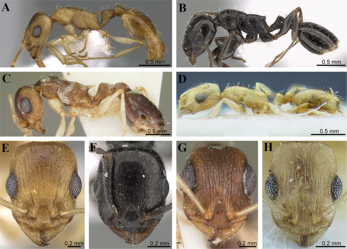
(A–H) Morphological overview of Arabian Temnothorax fauna. Body in profile and head in full face view. (A,E) Temnothorax arabicus Sharaf & Akbar (CASENT0746640 – AntWeb, photographer: Zach Lieberman); (B,F) Temnothorax elmenshawyi sp. n. (CASENT0922350 – AntWeb, photographer: Michele Esposito); (C,G) Temnothorax liviae (Agosti & Collingwood) (CASENT0102700 – AntWeb, photographer: April Nobile); (D,H) Temnothorax megalops (Hamann & Klemm) (CASENT0712601 – AntWeb, photographer: Matthew Prebus).

-
Eyes well developed but significantly smaller (OI 32–37) (Fig. 4A,B,D–F,H)……..2
-
2.
Body colour very dark brown to blackish; cephalic dorsum and sides of head behind posterior eye level mostly unsculptured and shiny with sparse punctate ground sculpture (Fig. 4B,F) ………………………………………………………………………………..…….T. elmenshawyi
-
Body colour yellowish; most of head with distinct rugose/rugulose sculpture (Fig. 4A,D,E,H)………………………………………………………………………………………………3
-
3.
Frontal carinae well-developed; propodeal spines long and acute; petiole with two pairs of erect setae (Fig. 4A,E)……………………………………………………………………………………T. arabicus
-
Frontal carinae feeble; propodeal spines short and blunt; petiole with a single pair of erect setae (Fig. 4D,H)…………………………………………………………………………………………………..T. megalops.
Discussion
This new species belongs to the T. exilis species group as defined by Cagniant & Espadaler12 in the inventory of Moroccan Temnothorax, and will not key successfully to any of the Moroccan or African species4. It has to be pointed out though, that the T. exilis species group is not a monophyletic group based on a recent global phylogeny of the genus5, which was already suspected by Cagniant & Espadaler12. Without any other existing higher-level classification above species level available for the genus, we prefer to place T. elmenshawyi in the T. exilis group, at least temporarily.
Temnothorax elmenshawyi superficially resembles T. exilis (Emery) from Italy but can be readily distinguished by the deeply impressed metanotal groove, the finely punctate posterior half of the cephalic surface, and absence of median clypeal carina. Workers of T. exilis have an indistinct metanotal groove, acute propodeal spines, a reticulate-rugulose posterior half of cephalic surface, and a median clypeal carina. With regards to the Arabian Temnothorax fauna, T. elmenshawyi cannot be confused with any of the Arabian species. It can be immediately separated by the uniformly very dark brown to blackish colour (vs. yellowish in the other three species), moderately large eyes (vs. extremely large eyes in T. liviae), and the mostly unsculptured posterior half of the head behind the level of the eyes (vs. conspicuously rugulose/rugose in the other three species).
More than 220 pitfall traps were placed in the Asir Mountains during two years of sampling and only two specimens of the new species were trapped. Undoubtedly, this may reflect the scarcity of this species and indicate a high probability of it being a regional endemic. Due to the extension of the Asir Mountains into Yemen, T. elmenshawyi may also occur in that country. It is hoped that future surveys of ants of this region will confirm this distribution, and result in the discovery of additional specimens, especially the male and queen castes.
As discussed in previous studies19,21, the use of micro-CT has great advantages for taxonomy. The non-destructive x-ray scan allows the generation of a virtual avatar or cybertype of the physical type material, providing a detailed and almost complete virtual reconstruction of the morphology, thus permitting a high degree of interactive examination for users without access to the physical specimen. Since standard ant taxonomy is still mostly based on the study and comparison of external morphology, virtual study of cybertype datasets can obviate the need to obtain loans from natural history museums, which are often difficult to organize, or to travel to the actual museum collection, which are usually costly and time-intensive. This is of special importance for developing countries where loans are almost never shipped to and travel budgets rather limited. The cybertype provided in this study is the first such virtual 3D dataset for an insect from the Arabian Peninsula or for the ant genus Temnothorax.
Material and Methods
Measurements and indices
The specimens were measured with Leica M205C stereomicroscope at magnifications of up to 160x. Measurements are expressed in millimeters and following24,25,26.
EL Eye length: maximum diameter of compound eye measured in oblique lateral view.
FRS Distance of the frontal carinae immediately caudal of posterior intersection points between frontal carinae and lamellae dorsal of torulus.
HL Head length: maximum distance from mid-point of anterior clypeal margin to mid-point of posterior margin of head, measured in full-face view.
HW Head width: width of head directly behind eyes, measured in full-face view.
IOcD Inter-ocellar distance: minimum distance between posterior-most pair of ocelli.
IOD Inter-ocular distance: minimum distance between compound eyes, measured in full-face view.
MD Malar distance: minimum distance between anterior margin of compound eye and base of mandible.
PH Petiole height: maximum height of petiole, measured from apex of node to ventral edge of petiole, parallel to anterior margin of petiole.
PL Petiole length: maximum length of petiole node measured in dorsal view from anterior notch close to propodeum to articulation with postpetiole.
PPH Postpetiole height: maximum height of postpetiole measured in lateral view from highest (median) point of node to ventral outline.
PPL Postpetiole length: maximum length of postpetiole node measured in dorsal view, excluding helcium.
PPW Postpetiole width: maximum width of postpetiole node measured in dorsal view.
PTW Petiole width: maximum width of petiole node measured in dorsal view.
PW Pronotal width: maximum width of pronotum measured in dorsal view.
SL Scape length: maximum scape length excluding basal condyle and neck.
SPST Distance between center of propodeal stigma and spine tip. The stigma center refers to midpoint defined by outer cuticular ring but not to center of stigma opening, which may be positioned eccentrically.
WL Weber’s length: diagonal length of mesosoma in lateral view from postero-ventral margin of propodeal lobe to anterior-most point of pronotal slope, excluding the neck.
Indices.
CI Cephalic index: HW/HL × 100
DPeI Dorsal petiole index: PTW/PTL × 100
DPpI Dorsal postpetiole index: PPW/PPL × 100
LPpI Lateral postpetiole index: PPL/PPH × 100
OI Ocular index: EL/HW × 100
PeNI Petiolar node index: PTW/PW × 100
PPI Postpetiole index: PPW/PTW × 100
PpNI Postpetiolar node index: PPW/PW × 100
PSLI Propodeal spine index: SPST/HL × 100
SI Scape index: SL/HL × 100
X-ray micro computed tomography and 3D images
The micro-CT scan was performed using a Zeiss Xradia 510 Versa 3D X-ray microscope operated with the Zeiss Scout-and-Scan Control System software (version 11.1.6411.17883). 3D reconstruction of the resulting scan raw data was done with the Zeiss Scout-and- Scan Control System Reconstructor (version 11.1.6411.17883) and saved in DICOM file format. Volume renderings, surface mesh generations, and virtual examinations were performed with Amira software (version 6.3.0). Post-processing of mesh data in order to generate a clean surface was done with Meshlab (version 1.3.3). The methodology for the virtual examination of 3D surface models, generation of 3D rotation videos, and virtual dissections follow Hita Garcia et al.21. For further details on micro-CT scanning and post-processing workflow pipeline, we refer to Hita Garcia et al.21,23.
Data Availability
As in previous studies21,23, the specimens used in this study have been databased and the data are freely accessible on AntWeb (http://www.antweb.org). Each specimen can be traced by a unique specimen identifier attached to its pin. The Cybertype dataset has been archived and is freely available from the Dryad Digital Repository (23, https://doi.org/10.5061/dryad.4gg39k6). In addition to the cybertype data at Dryad, we also provide a freely accessible 3D surface model on Sketchfab (https://skfb.ly/6HYRz). All data needed to evaluate the conclusions in the paper are present in the paper. Additional data related to this paper may be requested from the authors.
References
Mayr, G. Die Europäischen Formiciden. (Ameisen). Nach der analytischen Methode bearbeitet. Gerold, Vienna (1861).
Bolton, B. Afrotropical species of the myrmicine ant genera Cardiocondyla, Leptothorax, Melissotarsus, Messor and Cataulacus (Formicidae). Bull. Br. Mus. (Nat. Hist.). Entomol. 45, 307–370 (1982).
Bolton, B. An online catalog of the ants of the world. Available at http://antcat.org/[accessed 19 December 2017] (2017).
Prebus, M. Palearctic elements in the Old World tropics: a taxonomic revision of the ant genus Temnothorax Mayr (Hymenoptera, Formicidae) for the Afrotropical biogeographical region. ZooKeys 483, 23–57, https://doi.org/10.3897/zookeys.483.9111 (2015).
Prebus, M. Insights into the evolution, biogeography and natural history of the acorn ants, genus Temnothorax Mayr (Hymenoptera: Formicidae). BMC Evol. Biol. 17, 250, https://doi.org/10.1186/s12862-017-1095-8 (2017).
Guenard, B., Shik, J.Z., Booher, D., Lubertazzi D. & Alpert G. Extreme polygyny in the previously unstudied subtropical ant Temnothorax tuscaloosae (Hymenoptera: Formicidae), with implications for the biogeographic study of the evolution of polygyny. Ins. Soc. 63(4), https://doi.org/10.1007/s00040-016-0498-7 (2016).
Forel, A. Fourmis de Tunisie et de l’Algérie orientale. Ann. Soc. Entom. Belg. 34, lxi–lxxvi (1890).
Bernard, F. Faune de l’Europe et du Bassin Méditerranéen 3. Les fourmis d’Europe occidentale et septentrionale (1968): 411 pp. Paris (1967).
Bolton, B. Synopsis and classification of Formicidae. Mem. Am. Entomol. Inst. 71, 1–361 (2003).
Ward, P. S., Brady, S. G., Fisher, B. L. & Schultz, T. R. The evolution of myrmicine ants: phylogeny and biogeography of a hyperdiverse ant clade (Hymenoptera: Formicidae). Syst. Entom. 40, 61–81, https://doi.org/10.1111/syen.12090 (2015).
Fisher, B. L. & Bolton, B. Ants of Africa and Madagascar, A Guide to the Genera. Berkeley: University of California Press, 503 pp (2016).
Cagniant, H. & Espadaler, X. Les Leptothorax, Epimyrma et Chalepoxenus du Maroc (Hymenoptera: Formicidae). Clé et catalogue des espèces. Ann. Soc. Entom. Fr. 33, 259–284 (1997).
Hebert, P. D. N. & Gregory, T. R. The promise of DNA barcoding for taxonomy. Syst. Biol. 54, 852–859, https://doi.org/10.1080/10635150500354886 (2005).
Miller, S. E. DNA barcoding and the renaissance of taxonomy. PNAS 104, 4775–4776 (2007).
Giribet, G. A new dimension in combining data? The use of morphology and phylogenomic data in metazoan systematics. Acta Zool. 91, 11–9 (2010).
Ward, P. S. Integrating molecular phylogenetic results into ant taxonomy (Hymenoptera: Formicidae). Myrmecol. News 15, 21–9 (2011).
Csősz, S. & Fisher, B. L. Toward objective, morphology-based taxonomy: a case study on the Malagasy Nesomyrmex sikorai species group (Hymenoptera: Formicidae). PLoS ONE 11(4), e0152454, https://doi.org/10.1371/journal.pone.0152454. (2016).
Schlick-Steiner, B. C. et al. Integrative taxonomy: a multisource approach to exploring biodiversity. Annu Rev Entomol. 55, 421–438, https://doi.org/10.1146/annurev-ento-112408-085432 (2010).
Faulwetter, S., Vasileiadou, A., Kouratoras, M., Dailianis, T. & Arvanitidis, C. Micro-computed tomography: introducing new dimensions to taxonomy. Zookeys 263, 1–45, https://doi.org/10.3897/zookeys.263.4261 (2013).
Friedrich, F. et al. Insect morphology in the age of phylogenomics: innovative techniques and its future role in systematics. Entomol Sci. 17(1), 1–24, https://doi.org/10.1111/ens.12053 (2014).
Hita Garcia, F. et al. X-Ray microtomography for ant taxonomy: An exploration and case study with two new Terataner (Hymenoptera, Formicidae, Myrmicinae) species from Madagascar. PLoS ONE 12, e0172641, https://doi.org/10.1371/journal.pone.0172641 (2017).
Akkari, N., Enghoff, H. & Metscher, B. D. A new dimension in documenting new species: high-detail imaging for myriapod taxonomy and first 3D cybertype of a new millipede species (Diplopoda, Julida, Julidae). PLoS ONE. 10(8), e0135243, https://doi.org/10.1371/journal.pone.0135243 (2015).
Hita Garcia, F., Fischer, G., Liu, C., Audisio, T. L. & Economo, E. P. Next-generation morphological character discovery and evaluation: an X-ray micro-CT enhanced revision of the ant genus Zasphinctus Wheeler (Hymenoptera, Formicidae, Dorylinae) in the Afrotropics. ZooKeys 693, 33–93, https://doi.org/10.3897/zookeys.693.13012 (2017).
Sharaf, M. R., Akbar, S. A., Al Dhafer, H. M., El-Gharbawy, A. & Aldawood, A. S. Taxonomy of the Myrmicine ant genus Temnothorax Mayr, 1861 (Formicidae: Myrmicinae) in the Arabian Peninsula. Eur. J. of Tax. 280, 1–17, https://doi.org/10.5852/ejt.2017.280 (2017).
Snelling, R. R., Borowiec, M. L. & Prebus, M. M. Studies on California ants: a review of the genus Temnothorax. ZooKeys 372, 27–89, https://doi.org/10.3897/zookeys.372.6039 (2014).
Hita Garcia, F. & Fischer, G. Additions to the taxonomy of the Afrotropical Tetramorium weitzeckeri species complex (Hymenoptera, Formicidae, Myrmicinae), with the description of a new species from Kenya. Eur. J. of Tax. 90, 1–16, https://doi.org/10.5852/ejt.2014.90 (2014).
Acknowledgements
We are grateful to Brian Taylor, Matthew Prebus, Sandor Csosz, Henri Cagniant and Xavier Espadaler for useful discussions on the new species. Special thanks to Boris Kondratieff for useful suggestions. We thank Brian Fisher and Michele Esposito for imaging the new species and Ahmed Shams Al ‘Ola for photographing the type locality. This work was supported by the Deanship of Scientific Research at King Saud University [RG-1438-010]. EPE and FHG were supported by subsidy funding to OIST and JSPS Kakenhi Grants-in-Aid (No. 17K15180 to EPE, No. 18K14768 to FHG).
Author information
Authors and Affiliations
Contributions
M.R.S., F.H.G., E.P.E., A.A.W., A.S.A., conceived and designed the experiments, performed the practical work, analyzed the data, contributed analysis tools. M.R.S., F.H.G. prepared figures. E.P.E. also reviewed all drafts of the ms.
Corresponding author
Ethics declarations
Competing Interests
The authors declare no competing interests.
Additional information
Publisher’s note: Springer Nature remains neutral with regard to jurisdictional claims in published maps and institutional affiliations.
Rights and permissions
Open Access This article is licensed under a Creative Commons Attribution 4.0 International License, which permits use, sharing, adaptation, distribution and reproduction in any medium or format, as long as you give appropriate credit to the original author(s) and the source, provide a link to the Creative Commons license, and indicate if changes were made. The images or other third party material in this article are included in the article’s Creative Commons license, unless indicated otherwise in a credit line to the material. If material is not included in the article’s Creative Commons license and your intended use is not permitted by statutory regulation or exceeds the permitted use, you will need to obtain permission directly from the copyright holder. To view a copy of this license, visit http://creativecommons.org/licenses/by/4.0/.
About this article

Cite this article
Sharaf, M.R., Aldawood, A.S., Economo, E.P. et al. Taxonomy of Arabian Temnothorax Mayr (Formicidae: Myrmicinae) with description of a new species enhanced by x-ray microtomography. Sci Rep 9, 11009 (2019). https://doi.org/10.1038/s41598-019-47260-y
Received:
Accepted:
Published:
DOI: https://doi.org/10.1038/s41598-019-47260-y
Comments
By submitting a comment you agree to abide by our Terms and Community Guidelines. If you find something abusive or that does not comply with our terms or guidelines please flag it as inappropriate.
