Abstract
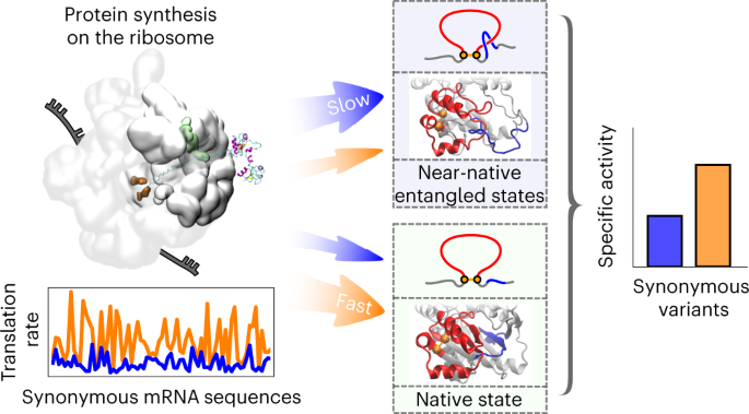
The specific activity of enzymes can be altered over long timescales in cells by synonymous mutations that alter a messenger RNA molecule’s sequence but not the encoded protein’s primary structure. How this happens at the molecular level is unknown. Here, we use multiscale modelling of three Escherichia coli enzymes (type III chloramphenicol acetyltransferase, d-alanine–d-alanine ligase B and dihydrofolate reductase) to understand experimentally measured changes in specific activity due to synonymous mutations. The modelling involves coarse-grained simulations of protein synthesis and post-translational behaviour, all-atom simulations to test robustness and quantum mechanics/molecular mechanics calculations to characterize enzymatic function. We show that changes in codon translation rates induced by synonymous mutations cause shifts in co-translational and post-translational folding pathways that kinetically partition molecules into subpopulations that very slowly interconvert to the native, functional state. Structurally, these states resemble the native state, with localized misfolding near the active sites of the enzymes. These long-lived states exhibit reduced catalytic activity, as shown by their increased activation energies for the reactions they catalyse.

This is a preview of subscription content, access via your institution
Access options
Access Nature and 54 other Nature Portfolio journals
Get Nature+, our best-value online-access subscription
$29.99 / 30 days
cancel any time
Subscribe to this journal
Receive 12 print issues and online access
$259.00 per year
only $21.58 per issue
Buy this article
- Purchase on Springer Link
- Instant access to full article PDF
Prices may be subject to local taxes which are calculated during checkout


Data availability
Data supporting the main findings of this study are available within the Article and its Supplementary Information and source data files. We cannot feasibly provide all ~5.3 TB of molecular dynamics trajectory data, but we provide the input data that were used to perform the simulations in this study in the repository subdirectory https://github.com/obrien-lab/cg_simtk_protein_folding/blob/master/example/input_data.tar.xz. All of the data that support the findings of this study, as well as the biological materials that were used to test the enzymatic activity of the DDLB and DHFR variants and for the LiP-MS experiments, are available from the corresponding author upon reasonable request. The raw mass spectrometry data for DDLB and DHFR have been deposited to the ProteomeXchange Consortium via the PRIDE partner repository with the dataset identifier PXD031425. A website (https://obrien-lab.github.io/visualize_entanglements/) was created to provide interactive visualization of the key misfolded, entangled structures predicted in this study. Source data are provided with this paper.
Code availability
All of the computer code developed in this work is available in the GitHub repositories https://github.com/obrien-lab/cg_simtk_protein_folding and https://github.com/obrien-lab/Activation-Energy-Estimation-Workflow under the MIT License. Detailed instructions on code usage, basic theory and examples of the input/output are available in the wiki pages of the above repositories.
References
Komar, A. A., Lesnik, T. & Reiss, C. Synonymous codon substitutions affect ribosome traffic and protein folding during in vitro translation. FEBS Lett. 462, 387–391 (1999).
Zhao, F., Yu, C.-H. & Liu, Y. Codon usage regulates protein structure and function by affecting translation elongation speed in Drosophila cells. Nucleic Acids Res. 45, 8484–8492 (2017).
Spencer, P. S., Siller, E., Anderson, J. F. & Barral, J. M. Silent substitutions predictably alter translation elongation rates and protein folding efficiencies. J. Mol. Biol. 422, 328–335 (2012).
Hunt, R. et al. A single synonymous variant (c.354G>A [p.P118P]) in ADAMTS13 confers enhanced specific activity. Int. J. Mol. Sci. 20, 5734 (2019).
Crombie, T., Boyle, J. P., Coggins, J. R. & Brown, A. J. The folding of the bifunctional TRP3 protein in yeast is influenced by a translational pause which lies in a region of structural divergence with Escherichia coli indoleglycerol‐phosphate synthase. Eur. J. Biochem. 226, 657–664 (1994).
Walsh, I. M. Testing the Effects of Synonymous Codon Usage on Co-Translational Protein Folding Using Novel Experimental and Computational Techniques. PhD thesis, Univ. Notre Dame (2019).
Yu, C.-H. et al. Codon usage influences the local rate of translation elongation to regulate co-translational protein folding. Mol. Cell 59, 744–754 (2015).
Walsh, I. M., Bowman, M. A., Santarriaga, I. F. S., Rodriguez, A. & Clark, P. L. Synonymous codon substitutions perturb cotranslational protein folding in vivo and impair cell fitness. Proc. Natl Acad. Sci. USA 117, 3528–3534 (2020).
Sala, A. J., Bott, L. C. & Morimoto, R. I. Shaping proteostasis at the cellular, tissue, and organismal level. J. Cell Biol. 216, 1231–1241 (2017).
Liu, Y. et al. Small molecule probes to quantify the functional fraction of a specific protein in a cell with minimal folding equilibrium shifts. Proc. Natl Acad. Sci. USA 111, 4449–4454 (2014).
Buhr, F. et al. Synonymous codons direct cotranslational folding toward different protein conformations. Mol. Cell 61, 341–351 (2016).
Martelli, P. L., Fariselli, P. & Casadio, R. Prediction of disulfide-bonded cysteines in proteomes with a hidden neural network. Proteomics 4, 1665–1671 (2004).
Niemyska, W. et al. Complex lasso: new entangled motifs in proteins. Sci. Rep. 6, 36895 (2016).
Sulkowska, J. I. On folding of entangled proteins: knots, lassos, links and θ-curves. Curr. Opin. Struct. Biol. 60, 131–141 (2020).
Baiesi, M., Orlandini, E., Seno, F. & Trovato, A. Sequence and structural patterns detected in entangled proteins reveal the importance of co-translational folding. Sci. Rep. 9, 8426 (2019).
Baiesi, M., Orlandini, E., Seno, F. & Trovato, A. Exploring the correlation between the folding rates of proteins and the entanglement of their native states. J. Phys. A: Math. Theor. 50, 504001 (2017).
Connolly, M. L., Kuntz, I. & Crippen, G. M. Linked and threaded loops in proteins. Biopolymers 19, 1167–1182 (1980).
Jarmolinska, A. I., Gambin, A. & Sulkowska, J. I. Knot_pull—python package for biopolymer smoothing and knot detection. Bioinformatics 36, 953–955 (2020).
Jennings, P. A., Finn, B. E., Jones, B. E. & Matthews, C. R. A reexamination of the folding mechanism of dihydrofolate reductase from Escherichia coli: verification and refinement of a four-channel model. Biochemistry 32, 3783–3789 (1993).
Garbuzynskiy, S. O., Ivankov, D. N., Bogatyreva, N. S. & Finkelstein, A. V. Golden triangle for folding rates of globular proteins. Proc. Natl Acad. Sci. USA 110, 147–150 (2013).
Nissley, D. A. et al. Universal protein misfolding intermediates can bypass the proteostasis network and remain soluble and less functional. Nat. Commun. 13, 3081 (2022).
Feng, Y. et al. Global analysis of protein structural changes in complex proteomes. Nat. Biotechnol. 32, 1036–1044 (2014).
Kröger, M. Developments in polymer theory and simulation. Polymers (Basel) 12, 30 (2019).
Pawlak, A. The entanglements of macromolecules and their influence on the properties of polymers. Macromol. Chem. Phys. 220, 1900043 (2019).
Sułkowska, J. I., Sułkowski, P. & Onuchic, J. Dodging the crisis of folding proteins with knots. Proc. Natl Acad. Sci. USA 106, 3119–3124 (2009).
Haglund, E. et al. Pierced lasso bundles are a new class of knot-like motifs. PLoS Comput. Biol. 10, e1003613 (2014).
Haglund, E. et al. The unique cysteine knot regulates the pleotropic hormone leptin. PLoS ONE 7, e45654 (2012).
Lu, H. P., Xun, L. & Xie, X. S. Single-molecule enzymatic dynamics. Science 282, 1877–1882 (1998).
Yang, H. et al. Protein conformational dynamics probed by single-molecule electron transfer. Science 302, 262–266 (2003).
Heidary, D. K., O’Neill, J. C., Roy, M. & Jennings, P. A. An essential intermediate in the folding of dihydrofolate reductase. Proc. Natl Acad. Sci. USA 97, 5866–5870 (2000).
Bitran, A., Jacobs, W. M., Zhai, X. & Shakhnovich, E. Cotranslational folding allows misfolding-prone proteins to circumvent deep kinetic traps. Proc. Natl Acad. Sci. USA 117, 1485–1495 (2020).
Towns, J. et al. XSEDE: accelerating scientific discovery. Comput. Sci. Eng. 16, 62–74 (2014).
O’Brien, E. P., Christodoulou, J., Vendruscolo, M. & Dobson, C. M. Trigger factor slows co-translational folding through kinetic trapping while sterically protecting the nascent chain from aberrant cytosolic interactions. J. Am. Chem. Soc. 134, 10920–10932 (2012).
Sharma, A. K., Bukau, B. & O’Brien, E. P. Physical origins of codon positions that strongly influence cotranslational folding: a framework for controlling nascent-protein folding. J. Am. Chem. Soc. 138, 1180–1195 (2016).
Fritch, B. et al. Origins of the mechanochemical coupling of peptide bond formation to protein synthesis. J. Am. Chem. Soc. 140, 5077–5087 (2018).
Nissley, D. A. & O’Brien, E. P. Structural origins of FRET-observed nascent chain compaction on the ribosome. J. Phys. Chem. B 122, 9927–9937 (2018).
Leininger, S. E., Trovato, F., Nissley, D. A. & O’Brien, E. P. Domain topology, stability, and translation speed determine mechanical force generation on the ribosome. Proc. Natl Acad. Sci. USA 116, 5523–5532 (2019).
Nissley, D. A. et al. Electrostatic interactions govern extreme nascent protein ejection times from ribosomes and can delay ribosome recycling. J. Am. Chem. Soc. 142, 6103–6110 (2020).
Dunkle, J. A. et al. Structures of the bacterial ribosome in classical and hybrid states of tRNA binding. Science 332, 981–984 (2011).
Arenz, S. et al. A combined cryo-EM and molecular dynamics approach reveals the mechanism of ErmBL-mediated translation arrest. Nat. Commun. 7, 12026 (2016).
Sharma, A. K. et al. A chemical kinetic basis for measuring translation initiation and elongation rates from ribosome profiling data. PLoS Comput. Biol. 15, e1007070 (2019).
Fluitt, A., Pienaar, E. & Viljoen, H. Ribosome kinetics and aa–tRNA competition determine rate and fidelity of peptide synthesis. Comput. Biol. Chem. 31, 335–346 (2007).
Eastman, P. et al. OpenMM 7: rapid development of high performance algorithms for molecular dynamics. PLoS Comput. Biol. 13, e1005659 (2017).
Nagano, N. EzCatDB: the enzyme catalytic-mechanism database. Nucleic Acids Res. 33, D407–D412 (2005).
Nagano, N. et al. EzCatDB: the enzyme reaction database, 2015 update. Nucleic Acids Res. 43, D453–D458 (2014).
The UniProt Consortium. UniProt: the universal protein knowledgebase. Nucleic Acids Res. 46, 2699 (2018).
Berman, H. M. et al. The Protein Data Bank. Nucleic Acids Res. 28, 235–242 (2000).
Acknowledgements
S.D.F. acknowledges support from the National Institutes of Health (NIH) Director’s New Innovator Award (DP2GM140926) and National Science Foundation (MCB-2045844). S.J.B. acknowledges support from the NIH (GM-122595), Eberly Family Distinguished Chair in Science and Howard Hughes Medical Institute. E.P.O. acknowledges support from the National Science Foundation (MCB-1553291) and NIH (R35-GM124818). Computations in this work were carried out on the Extreme Science and Engineering Discovery Environment supercomputer32 (which is supported by MCB-160069) and the Pennsylvania State University’s Institute for Computational and Data Sciences’ Roar supercomputer. The CLS Behring Fermentation Facility and Huck Institutes of the Life Sciences at the Pennsylvania State University provided equipment and training to grow and purify DHFR biological replicates. We thank T. Berek and P. Kashyap for help with growing and purifying the DHFR biological replicates.
Author information
Authors and Affiliations
Contributions
E.P.O. designed the research. Y.J. developed the computational methods with contributions from E.P.O. Y.J. wrote the computer code and carried out the simulations and computations. S.S.N. and S.J.B. designed the experimental validation for the DDLB variants. S.S.N., I.S., E.P.O. and S.J.B. designed the experimental validation for the DHFR variants. S.S.N., I.S. and P.P. performed the specific activity experiments. S.D.F. designed the LiP-MS experiments for DDLB and DHFR. P.T. and Y.X. performed the LiP-MS experiments. All of the authors analysed the data and wrote the manuscript.
Corresponding author
Ethics declarations
Competing interests
The authors declare no competing interests.
Peer review
Peer review information
Nature Chemistry thanks Kevin Pagel and the other, anonymous, reviewer(s) for their contribution to the peer review of this work.
Additional information
Publisher’s note Springer Nature remains neutral with regard to jurisdictional claims in published maps and institutional affiliations.
Supplementary information
Supplementary Information
Supplementary Methods, Supplementary Results (virtual screening and disentangling timescale), Supplementary Discussions, Supplementary Tables 1–15 and Supplementary Figs. 1–15.
Supplementary Video 1
Co- and post-translational folding trajectory of a misfolded CAT-III with a non-native entanglement formed after synthesis. The conformation gets trapped in state P13 at the end. The nascent chain is shown in cyan, where the closed loop formed by the native contact is highlighted in red and the segment threading the red loop is highlighted in blue. At each nascent chain length, the conformation is obtained from the last frame of the simulation trajectory. The time of ejection, dissociation and post-translation processes are presented using the experimental timescale on the top left of the scene.
Supplementary Video 2
Co- and post-translational folding trajectory of a correctly folded CAT-III without any non-native entanglements formed. The nascent chain is coloured based on secondary structure elements, where α helices are magenta and β sheets are yellow. At each nascent chain length, the conformation is obtained from the last frame of the simulation trajectory. The time of ejection, dissociation and post-translation processes are presented using the experimental timescale on the top left of the scene.
Supplementary Video 3
Co- and post-translational folding trajectory of a misfolded DDLB with a non-native entanglement formed during synthesis that persisted in the post-translational dynamics. The conformation gets trapped in state P4 at the end. The nascent chain is shown in cyan, where the closed loop formed by the native contact is highlighted in red and the segment threading the red loop is highlighted in blue. At each nascent chain length, the conformation is obtained from the last frame of the simulation trajectory. The time of ejection, dissociation and post-translation processes are presented using the experimental timescale on the top left of the scene.
Supplementary Video 4
Co- and post-translational folding trajectory of a correctly folded DDLB without any non-native entanglements formed. The nascent chain is coloured based on secondary structure elements, where α helices are magenta and β sheets are yellow. At each nascent chain length, the conformation is obtained from the last frame of the simulation trajectory. The time of ejection, dissociation and post-translation processes are presented using the experimental timescale on the top left of the scene.
Source data
Source Data Fig. 1
Data presented in the figure and raw data points for statistics.
Source Data Fig. 2
Data presented in the figure and raw data points for statistics.
Source Data Fig. 3
Data presented in the figure and raw data points for statistics.
Source Data Fig. 4
Data presented in the figure and raw data points for statistics.
Source Data Fig. 5
Data presented in the figure.
Source Data Fig. 6
Raw pathway probabilities.
Rights and permissions
Springer Nature or its licensor (e.g. a society or other partner) holds exclusive rights to this article under a publishing agreement with the author(s) or other rightsholder(s); author self-archiving of the accepted manuscript version of this article is solely governed by the terms of such publishing agreement and applicable law.
About this article
Cite this article
Jiang, Y., Neti, S.S., Sitarik, I. et al. How synonymous mutations alter enzyme structure and function over long timescales. Nat. Chem. 15, 308–318 (2023). https://doi.org/10.1038/s41557-022-01091-z
Received:
Accepted:
Published:
Issue Date:
DOI: https://doi.org/10.1038/s41557-022-01091-z
This article is cited by
-
Codon language embeddings provide strong signals for use in protein engineering
Nature Machine Intelligence (2024)
-
Oxidative stress resistance prompts pyrroloquinoline quinone biosynthesis in Hyphomicrobium denitrificans H4-45
Applied Microbiology and Biotechnology (2024)
-
In silico methods for predicting functional synonymous variants
Genome Biology (2023)
-
Analysis of codon usage bias of thioredoxin in apicomplexan protozoa
Parasites & Vectors (2023)
-
Whisperings from not so silent mutations
Nature Reviews Microbiology (2023)


