Abstract
In tasks involving the interpretation of medical images, suitably trained machine-learning models often exceed the performance of medical experts. Yet such a high-level of performance typically requires that the models be trained with relevant datasets that have been painstakingly annotated by experts. Here we show that a self-supervised model trained on chest X-ray images that lack explicit annotations performs pathology-classification tasks with accuracies comparable to those of radiologists. On an external validation dataset of chest X-rays, the self-supervised model outperformed a fully supervised model in the detection of three pathologies (out of eight), and the performance generalized to pathologies that were not explicitly annotated for model training, to multiple image-interpretation tasks and to datasets from multiple institutions.
Main
Deep learning has enabled the automation of complex medical image interpretation tasks, such as disease diagnosis, often matching or exceeding the performance of medical experts1,2,3,4,5. However, despite these meaningful improvements in diagnostic efficiency, automated deep learning models often require large labelled datasets during training6. These large-scale labelling efforts can be expensive and time consuming, often requiring extensive domain knowledge or technical expertise to implement for a particular medical task7,8.
Several approaches such as model pre-training and self-supervision have been proposed to decrease model reliance on large labelled datasets9,10,11,12. Although self-supervised pre-training approaches have been shown to increase label efficiency across several medical tasks, they still require a supervised fine-tuning step after pre-training that requires manually labelled data for the model to predict relevant pathologies13,14. As a result, these approaches are only able to predict diseases that were explicitly annotated in the dataset, and are unable to predict pathologies that were not explicitly annotated for training. Thus, for the model to predict a certain pathology with reasonable performance, it must be provided with a substantial number of expert-labelled training examples for that pathology during training. This process of obtaining high-quality annotations of certain pathologies is often costly and time consuming, often resulting in large-scale inefficiencies in clinical artificial intelligence workflows.
In this Article, to address these limitations, we applied a machine-learning paradigm where a model can classify samples during test time that were not explicitly annotated during training15,16. We present a zero-shot method using a fully self-supervised-learning procedure that does not require explicit manual or annotated labels for chest X-ray image interpretation to create a model with high performance for the multi-label classification of chest X-ray images. The method, which we call CheXzero, uses contrastive learning, a type of self-supervised learning, with image–text pairs to learn a representation that enables zero-shot multi-label classification. The method can also be considered as a form of natural-language supervision or unsupervised learning15. In contrast to previous self-supervised approaches, the method does not require fine-tuning using labelled data. Hence, unlike previous self-supervised approaches, the method requires no labels except for testing, and is able to accurately identify pathologies that were not explicitly annotated. To develop the method, we leveraged the fact that radiology images are naturally labelled through corresponding clinical reports and that these reports can offer a natural source of supervision. We show that the performance of the self-supervised method is comparable to the performance of both expert radiologists and fully supervised methods on unseen pathologies in two independent test datasets collected from two different countries. We also show that the self-supervised method outperforms previous label-efficient approaches on chest X-ray pathology classification, suggesting that explicit labels are not required to perform well on medical-image-interpretation tasks when corresponding reports are available for training. Using chest X-rays as a driving example, the self-supervised method exemplifies the potential of deep-learning methods for learning a broad range of medical-image-interpretation tasks from large amounts of unlabelled data, thereby decreasing inefficiencies in medical machine-learning workflows that result from large-scale labelling efforts.
Results
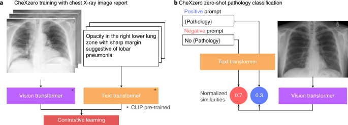
We leverage zero-shot learning to classify pathologies in chest X-rays without training on explicit labels (Fig. 1). To do so, we took image–text pairs of chest X-rays and radiology reports, and the model learned to predict which chest X-ray corresponds to which radiology report. We trained the model with 377,110 pairs of a chest X-ray image and the corresponding raw radiology report from the MIMIC-CXR dataset17.
a, Training pipeline. The model learns features from raw radiology reports, which act as a natural source of supervision. b, Prediction of pathologies in a chest X-ray image. For each pathology, we generated a positive and negative prompt (such as ‘consolidation’ versus ‘no consolidation’). By comparing the model output for the positive and negative prompts, the self-supervised method computes a probability score for the pathology, and this can be used to classify its presence in the chest X-ray image.
The performance of the self-supervised model is comparable to that of three benchmark radiologists classifying the five CheXpert competition pathologies evaluated on the CheXpert test dataset. On the Matthews correlation coefficient (MCC) metric, there is no statistically significant difference (model − radiologist performance = −0.005; 95% confidence interval (CI) −0.043, 0.034) between the performance of the model (0.523; 95% CI 0.486, 0.561) and that of the radiologists (0.530; 95% CI 0.499, 0.558) averaged over the pathologies. On individual pathologies, the model’s MCC performance is higher, but not statistically significantly, compared with radiologists on consolidation (0.018; 95% CI −0.090, 0.123), cardiomegaly (0.058; 95% CI −0.016, 0.133) and oedema (0.015; 95% CI −0.070, 0.099). The model’s MCC performance is lower, but not statistically significantly, compared with radiologists on atelectasis (−0.078; 95% CI −0.154, 0.000) and pleural effusion (−0.040; 95% CI −0.096, 0.013). On the F1 metric, there is similarly no statistically significant difference (model − radiologist performance = −0.009; 95% CI −0.038, 0.018) between the mean F1 performance of the model (0.606; 95% CI 0.571, 0.638) and that of the radiologists (0.619; 95% CI 0.585, 0.642) averaged over the pathologies. On individual pathologies, we find that the model F1 performance is significantly higher than that of radiologists on cardiomegaly (model − radiologist performance = 0.065; 95% CI 0.013, 0.115). We find that the model’s F1 performance is significantly lower than that of radiologists on atelectasis (model − radiologist performance = −0.045; 95% CI −0.090, −0.001). There are no statistically significant differences in F1 for consolidation (model − radiologist performance = −0.050; 95% CI −0.146, 0.036), oedema (model − radiologist performance = 0.018; 95% CI −0.053, 0.086) and pleural effusion (model − radiologist performance = −0.034; 95% CI −0.078, 0.008). Figure 2 shows the receiver operating characteristic (ROC) curve performance of the model and the radiologist operating points. Table 1 lists the mean performance of the radiologists and the model, and their associated difference with 95% CI.
a, F1 scores of the self-supervised model as compared with three board-certified radiologists on the CheXpert test dataset for the five CheXpert competition conditions. The model’s F1 score is significantly higher than that of radiologists on pleural effusion, significantly lower on atelectasis and not statistically significantly different on cardiomegaly, consolidation and oedema. b, Comparison of the MCC of the self-supervised model against three board-certified radiologists on the CheXpert test dataset. The MCC of the model is not significantly different than that of radiologists on all five pathologies. a,b, Green plots indicate the performance of the three board-certified radiologists while blue plots indicate the performance of the self-supervised model. c, Comparison of the ROC curve of the self-supervised model to benchmark radiologists against the test-set ground truth. The model outperforms the radiologists when the ROC curve lies above the radiologists’ operating points. The dotted lines on the ROC curves represent the baseline performance of a classifier that is no better than random guessing.
The results show that the self-supervised model outperforms three previous label-efficient methods (MoCo-CXR, MedAug and ConVIRT) on the CheXpert dataset, using no explicit labels during training. MoCo-CXR and MedAug use self-supervision using only chest X-ray images. Specifically, MoCo-CXR modifies the contrastive learning framework Momentum Contrast (MoCo) for chest X-ray interpretation. MedAug builds on MoCo pre-training by using patient metadata to select positive chest X-ray image pairs for image–image contrastive pre-training. ConVIRT uses chest X-rays along with associated report data to conduct self-supervision. Specifically, ConVIRT jointly trains a ResNet-50 and a Transformer by leveraging randomly sampled text from paired chest X-ray and radiology-report data to learn visual representations. Unlike our approach, these previous works require a small fraction of labelled data to enable pathology classification. The self-supervised model’s mean area under the curve (AUC) of 0.889 outperforms ConVIRT trained on 1% of the labelled data (AUC 0.870), ConVIRT trained on 10% of the labelled data (AUC 0.881), ConVIRT trained on 100% of the labelled data (AUC 0.881), MedAug trained on 1% of the labelled data (AUC 0.810), MoCo-CXR trained on 1% of the labelled data (AUC 0.802), MoCo-CXR trained on 10% of the labelled data (AUC 0.850) and MoCo-CXR trained on 100% of the labelled data (AUC 0.884) (Table 2). Additionally, on the task of classifying plural effusion, the self-supervised model’s mean AUC of 0.932 outperforms MoCo-CXR trained on 0.1% of the labelled data (AUC 0.813) and MoCo-CXR trained on 1% of the labelled data (AUC 0.885), MoCo-CXR trained on 10% of the labelled data (AUC 0.920) and MedAug trained on 1% of the labelled data (AUC 0.906) (Table 3)13,18. However, the self-supervised model achieves these results without the use of any labels or fine-tuning, thus showing the capability of the model on a zero-shot task.
The flexibility of zero-shot learning enables the self-supervised model to perform auxiliary tasks related to the content found in radiology reports. We applied the self-supervised model to tasks including differential diagnosis using the PadChest dataset, patient sex prediction and chest radiograph projection (anteroposterior versus posteroanterior) prediction19.
On the task of differential diagnosis on the PadChest dataset, we find that the model achieves an AUC of at least 0.900 on 6 findings and at least 0.700 on 38 findings out of 57 radiographic findings where n > 50 in the PadChest test dataset (n = 39,053). We obtain high performance on the CheXpert competition pathologies such as pleural effusion, oedema, atelectasis, consolidation and cardiomegaly, with AUCs of 0.958 (95% CI 0.953, 0.963) for pleural effusion, 0.961 (95% CI 0.946, 0.974) for oedema, 0.798 (95% CI 0.780, 0.817) for atelectasis, 0.871 (95% CI 0.851, 0.888) for consolidation and 0.898 (95% CI 0.894, 0.903) for cardiomegaly (Fig. 3). Compared with the performance of the CheXNet model on the PadChest dataset, we observe that the self-supervised model outperformed their approach on three out of the eight selected pathologies, atelectasis, consolidation and oedema, despite using 0% of the labels as compared with 100% in the CheXNet study (Table 4)20,21.
Mean AUC and 95% CI are shown for each radiographic finding (n > 50) labelled as high importance by an expert radiologist. We externally validated the model’s ability to generalize to different data distributions by evaluating model performance on the human-annotated subset of the PadChest dataset (n = 39,053 chest X-rays). No labelled samples were seen during training for any of the radiographic findings in this dataset. The self-supervised method achieves an AUC of at least 0.900 on 6 findings and at least 0.700 on 38 findings out of 57 radiographic findings where n > 50 in the PadChest test dataset (n = 39,053).
In addition to the ensembled self-supervised model, we trained a single model using full radiology reports instead of only the impressions section in order to evaluate zero-shot performance on auxiliary tasks such as the prediction of sex. The model trained with full radiology reports achieved an AUC of 0.936 (95% CI 0.910, 0.959) on sex prediction using the prompts ‘the patient’s sex is male’ and ‘the patient’s sex is female’. Additionally, the model achieved an AUC of 0.799 (95% CI 0.7595, 0.835) on the task of predicting whether a chest X-ray is anteroposterior or posteroanterior. To make these predictions on an auxiliary task, the model requires only the development of prompts to use for the task; no training or labels are needed.
Discussion
The purpose of this work was to develop and demonstrate performance of a zero-shot classification method for medical imaging without training on any explicit manual or annotated labels. The results show that, with no explicit labels, the zero-shot method is comparable to the performance of both expert radiologists and fully supervised methods on pathologies that were not explicitly labelled during training. Specifically, the self-supervised method achieved an AUC −0.042 points below that of the highest-performing fully supervised model on the CheXpert competition. We also show that the performance of the self-supervised model is comparable to that of radiologists, as there is no statistically significant difference between the performance of the model and the performance of the radiologists on the average MCC and F1 over the five CheXpert competition pathologies. We also show that the self-supervised model outperforms previous label-efficient approaches on chest X-ray pathology classification, suggesting that explicit labels are not required to perform well on medical-image-interpretation tasks when corresponding reports are available for training. We achieved these results using a deep-learning model that learns chest X-ray image features using corresponding clinically available radiology reports as a natural signal. In addition, we show that ensembling over the top-ten highest-performing model checkpoints on the validation dataset can improve the performance of the model (Table 5). We externally validated the self-supervised model, trained on the MIMIC-CXR dataset, on two independent datasets, the CheXpert test dataset and the human-annotated subset of the PadChest dataset.
Previous efforts for learning with small amounts of labelled data have shown meaningful improvements in performance using fewer labels, but still require the availability of some annotations that may not be trivial to obtain. For instance, recent work has achieved a mean AUC of 0.870 on the CheXpert test dataset using only 1% of the labelled data14. However, labelling 1% of a large dataset can still be expensive. For example, 1% of the labelled data in the ChestX-ray14, PadChest and CheXpert datasets amounts to 1,000 labels, 1,609 labels and 2,243 labels, respectively8,19. Additionally, these methods can only predict pathologies that were labelled during training, thereby restricting their applicability to other chest pathologies or classification tasks. Therefore, previous label-efficient learning methods may not be as potent in settings where access to a diverse set of high-quality annotations is limited. In contrast, the self-supervised method that we report in this work achieves a mean AUC of 0.889 on the CheXpert test dataset without requiring any explicit annotations (Tables 1 and 2). Recent work has leveraged radiology reports for zero-shot chest X-ray classification; however, it is applicable only to chest X-ray images with only one pathology, limiting the practicality of the method since multiple pathologies are often present in real-world settings22. Additionally, recent work has shown that a zero-shot learning approach can predict unseen chest X-ray pathologies, but the method still requires explicit labels during training23. Our model does not require labels for any pathology since we do not have to distinguish between ‘seen’ and ‘unseen’ classes during training.
To increase the number of labelled datasets and to reduce the effort required for manual annotations by domain experts, recent works have designed automatic labellers that can extract explicit labels from unstructured text reports. However, the development time of automatic labelling systems such as the NIH labeller and CheXpert are high, each requiring either extensive domain knowledge or technical expertise to implement7,24. This burden is not limited to chest X-rays; previous works have developed labelling methods for several forms of unstructured clinical text such as cancer-pathology reports and electronic health records25,26,27. In contrast, our method is able to classify pathologies without requiring the domain-specific development of an automatic labeller. The self-supervised method has the potential to alleviate the labelling bottleneck in the machine-learning pipeline for a range of medical-imaging tasks by leveraging easily accessible unstructured text data without domain-specific pre-processing efforts17. As a result, the self-supervised method opens promising avenues for approaches and applications in the medical-imaging domain, where narrative reports that describe imaging findings are common.
One notable finding is the ability of the self-supervised method to predict differential diagnoses and radiographic findings with high accuracy on a dataset that was collected in a country different from that of the training dataset19. This ability to generalize to datasets from vastly different distributions has been one of the primary challenges for the deployment of medical artificial intelligence28,29. Despite the challenges of generalization described in previous works, the self-supervised method achieves an AUC of at least 0.900 on 6 radiographic findings and at least 0.700 on 38 findings out of 57 radiographic findings where n > 50 in the PadChest test dataset (n = 39,053) (Fig. 3). We speculate that the self-supervised model can generalize better because of its ability to leverage unstructured text data, which contains more diverse radiographic information that could be applicable to other datasets. Additionally, we note that we might expect improved performance if we used alternative labels instead of the raw clinical findings in PadChest. Ultimately, the results demonstrate that the self-supervised method can generalize well on a different data distribution without having seen any explicitly labelled pathologies from PadChest during training30.
Biases may have affected the training of the self-supervised method. For example, if a pathology is never mentioned in the reports, then the method cannot be expected to predict that pathology with high accuracy during zero-shot evaluation. Furthermore, the model’s ability to predict a pathology may depend on the terminology used in the training reports. For instance, if several reports describe a condition such as atelectasis, but do not explicitly use the term, then the method may not perform well when queried with the phrase ‘has atelectasis’31. Thus, the method’s ability to predict pathologies is limited to scenarios mentioned in the text reports, and may perform less well when there are a variety of ways to describe the same pathology. To address these potential biases, we provide the model with hundreds of thousands of image–text pair samples (n = 377,110) during training, encompassing a wide variety of writing styles and descriptions of pathologies17. By validating the method on the CheXpert and PadChest datasets, which were collected at different hospitals from the one used in the training of the model, we show that site-specific biases are not inhibiting the method’s ability to predict clinically relevant pathologies with high accuracy.
This work has a few limitations. First, the self-supervised method still requires repeatedly querying performance on a labelled validation set for hyperparameter selection and to determine condition-specific probability thresholds when calculating MCC and F1 statistics. Second, the self-supervised method is currently limited to classifying image data; however, medical datasets often combine different imaging modalities, can incorporate non-imaging data from electronic health records or other sources, or can be a time series. For instance, magnetic resonance imaging and computed tomography produce three-dimensional data that have been used to train other machine-learning pipelines32,33,34. On the same note, it would be of interest to apply the method to other tasks in which medical data are paired with some form of unstructured text. For instance, the self-supervised method could leverage the availability of pathology reports that describe diagnoses such as cancer present in histopathology scans26,35,36. Lastly, future work should develop approaches to scale this method to larger image sizes to better classify smaller pathologies37,38,39,40,41,42,43,44,45.
In summary, we have designed a self-supervised method using contrastive learning that detects the presence of multiple pathologies in chest X-ray images. The self-supervised method builds on the use of image–text pairings of chest X-rays and radiology reports in ConVIRT, as well as on the multi-class zero-shot classification of natural images in Contrastive Language-Image Pre-training (CLIP) to enable the application of zero-shot approaches to medical-image interpretation. The self-supervised method matches radiologist-level performance on a chest X-ray classification task for multiple pathologies that the model was not explicitly trained to classify (Fig. 2 and Table 1). The results highlight the potential of deep-learning models to leverage large amounts of unlabelled data for a broad range of medical-image-interpretation tasks, and thereby may reduce the reliance on labelled datasets and decrease clinical-workflow inefficiencies resulting from large-scale labelling efforts.
Methods
Datasets
Training
The self-supervised method was trained on the MIMIC-CXR dataset, a publicly available dataset of chest radiographs with radiology text reports. The MIMIC-CXR dataset contains 377,110 images corresponding to 227,835 radiographic studies17. For instances where a radiographic study contains more than one chest X-ray image, the chest X-ray that is in anteroposterior/posteroanterior view was chosen to be included as part of training. Each radiographic study comes with a corresponding free-text radiology report, a summarization written by radiologists regarding their findings. Each full radiology report consists of multiple sections: examination, indication, impression, findings, technique and comparison. CheXpert is a public dataset for chest radiograph interpretation, consisting of 224,316 chest X-rays of 65,240 patients from Stanford Hospital8. The dataset is labelled for the presence of 14 different conditions: atelectasis, cardiomegaly, consolidation, oedema, enlarged cardiomediastinum, fracture, lung lesion, lung opacity, no finding, pleural effusion, pleural other, pneumonia, pneumothorax and support devices. These labels are obtained from the agreement of five board-certified radiologists. Additionally, the dataset consists of free-text radiology reports that are associated with each chest X-ray image. The CheXpert validation dataset is utilized for tuning-condition-specific probability thresholds to obtain predictions from the self-supervised model’s probabilities for the five CheXpert competition conditions of a given chest X-ray image We conduct this analysis by running inference with the self-supervised model to obtain probability values of each condition being present for all chest X-ray images. Condition-specific probability thresholds are then determined by choosing the probability values that result in the best MCC for each condition on the CheXpert validation dataset. The CheXpert validation dataset has no overlap with the CheXpert test dataset used for evaluation.
Evaluation
The self-supervised method was evaluated on two external datasets: the CheXpert test dataset and PadChest. The CheXpert test dataset is a collection of chest X-rays that are commonly used to evaluate the performance of models on chest X-ray interpretation tasks14,31. We evaluate the model on the entire CheXpert test dataset, consisting of 500 chest X-ray images labelled for the presence of 14 different conditions8. The CheXpert test dataset is utilized to calculate both the self-supervised model’s area under the receiver operating characteristic (AUROC) and MCC metrics for each of the five CheXpert competition conditions. Additionally, the test set contains predictions from three board-certified radiologists on full-resolution images with which we compare the performance of the model.
The PadChest dataset is a public dataset that contains 160,868 chest X-ray images labelled with 174 different radiographic findings, 19 differential diagnoses19. Twenty-seven per cent of the labels come from board-certified radiologists, and the rest were obtained by using a recurrent neural network with attention trained on the radiology reports. For evaluation purposes, only 39,053 examples from the dataset were utilized, each of which was annotated by board-certified radiologists. These examples were then used to calculate the self-supervised model’s AUROC for each of the different conditions described above.
Pre-processing
Each of the 377,110 chest X-rays in the MIMIC-CXR dataset were re-sized to 224 × 224 and zero padded before training. Each image was then normalized using a sample mean and standard deviation of the training dataset.
Text from radiology reports were tokenized using the byte pair encoding procedure with a vocabulary size of 49,408. For text that exceeds the maximum token sequence length of the given architecture, we truncated the text embedding to the first ‘context length tokens – 2’. The remaining two tokens were saved for the [SOS] and [EOS] tokens at the beginning and end of the text embedding, respectively.
Architecture
The uninitialized architectures consist of a Vision Transformer, ViT-B/32, for the image encoder, and a Transformer for the text encoder. We use a pre-trained Vision Transformer that accepts images of resolution 224 × 224. The text encoder Transformer has a base size of 63 million parameters, 12 layers and a width of 512 with 8 attention heads. The Transformer operates on lower-byte pair encoding representation of text and uses text embeddings with a maximum token length of 77. We use the same initialization scheme used in CLIP15.
Implementation of the method
Model pre-training
The self-supervised model consists of an image and text encoder that we jointly train on the MIMIC-CXR training dataset17. We utilize the impressions section of each text report, since it contains a concise summary of the entire report. We contrast this with a previous self-supervised method, ConVIRT, which selects a random sentence from the full-length radiology report for each image14. Although their proposed method could extract some signal, a random text input selection allows for unnecessary stochasticity that could lead to inconsistencies in training. To address this, we consistently select the text from the impressions section.
Training
We initialized the self-supervised model using the ViT-B/32and Transformer architectures with pre-trained weights from OpenAI’s CLIP model15. When training on the impressions section, we keep the maximum context length of 77 tokens as given in the CLIP architecture. We demonstrated that we can leverage the pre-trained weights from the CLIP architecture learned from natural images to train a zero-shot model with a domain-specific medical task.
To prepare the data for training, all images from the MIMIC-CXR dataset are stored in a single HDF5 file. We performed a hyperparameter sweep over the batch size and the learning rate using the CheXpert validation dataset. We compute the validation mean AUC over the five CheXpert competition pathologies after every 1,000 batches are trained, and save the model checkpoint if the model outperforms the last best model during training. The validation mean AUCs of these checkpoints are used to select models for ensembling. The best model uses stochastic gradient descent for optimization with a learning rate of 0.0001 and momentum of 0.9. The best model has a batch size of 64 and is trained for four epochs. We train the model by maximizing the cosine similarity between image and text embeddings of all valid image–report pairs in the batch while minimizing the cosine similarity between the embeddings of incorrect pairings in the batch. The method’s training procedure closely follows the implementation of CLIP15.
Softmax evaluation technique for multi-label classification
To evaluate the zero-shot performance of the model on the multi-label classification task, we used a positive–negative softmax evaluation procedure on each of the diseases. In contrast to CLIP, the proposed procedure allows us to normalize with respect to the negated version of the same disease classification instead of naively normalizing across the diseases to obtain probabilities from the logits15. The latter approach is less reasonable in this context since a single image may have multiple associated labels.
We define the procedure as follows. First, we compute logits with positive prompts (such as atelectasis) and negative prompts (that is, no atelectasis). Then, we compute the softmax between the positive and negative logits. Lastly, we keep the softmax probabilities of the positive logits as the probability that the disease is present in the chest X-ray.
Ensembling
We ensemble the top-ten model checkpoints sorted by mean AUC over the five CheXpert pathologies on the validation dataset. The probability outputs of the ensemble are computed by taking the average of the probability outputs of each model. The probabilities are averaged after softmax evaluation. These probabilities are then used for model evaluation through AUC and for prediction tasks using condition thresholds generated from the validation dataset.
Knowledge-distillation procedure
To allow for the use of the CLIP pre-trained model on full radiology reports to evaluate zero-shot performance on auxiliary tasks such as sex prediction, we use a knowledge-distillation procedure. This procedure is required as the pre-trained text encoder from the CLIP model has a context length of only 77 tokens, which is not long enough for an entire radiology report. We use the pre-trained model to train a model with a context length of 512, long enough to encompass 98% of radiology reports. In this method, the text encoder of the best-performing model trained only on impressions is used as a teacher for the text encoder of a student model. To train the student, we compute the mean squared error between the logits of the two encoders, then backpropagate across the student architecture. Once the student text encoder is trained, we replace the uninitialized image encoder in the student model with the image encoder of the teacher model. Then, the student model is contrastively trained on the MIMIC-CXR chest X-ray and full-text report pairs.
Prompt-engineering methods
We run experiments using the labels present in the test set as the prompts and creating the prompts of ‘<label>’ and ‘no <label>’ as the positive and negative prompts for the softmax evaluation procedure.
Statistical analysis
AUROC
We collect AUROC results from both the CheXpert test dataset (500 samples) as well as PadChest dataset (39,053 samples) using the self-supervised model’s predictions. The AUROC and MCC results of the five clinically relevant pathologies on the CheXpert test dataset are presented in Table 1. Table 2 consists of the mean AUROC of these five pathologies on the CheXpert test dataset along with self-supervised and supervised comparisons. The DAM supervised method is included as a comparison and currently is state-of-the-art on the CheXpert dataset. An additional supervised baseline, DenseNet121, trained on the CheXpert dataset is included as a comparison since DenseNet121 is commonly used in self-supervised approaches. Current top-performing label-efficient approaches, ConVIRT, MedAug and MoCo-CXR, are included as self-supervised comparisons.
MCC and F1 score
To obtain the MCC, we first run inference on the CheXpert test set using our softmax evaluation technique to obtain probability values for the 14 different conditions on each of the 500 chest X-ray images. The probabilities are then transformed into positive/negative predictions using the probability thresholds computed by optimizing MCC over the validation dataset. Then, the condition-based MCC scores are calculated using these predictions. We similarly compute the F1 score, but using the same thresholds as used for computing the MCC.
Confidence intervals
We use the non-parametric bootstrap to generate confidence intervals: random samples of size n (equal to the size of the original dataset) are repeatedly sampled 1,000 times from the original dataset with replacement. We then estimate the AUROC, F1 and MCC metrics (or their difference for two the methods) using each bootstrap sample. We derive confidence intervals from the relative frequency distribution of the estimates over the re-samples, using the interval between the 100 × (α/2) and 100 × (1 − α/2) percentiles; we pick α = 0.05.
Reporting summary
Further information on research design is available in the Nature Research Reporting Summary linked to this article.
Data availability
The main data (CheXpert data) supporting the results of this study are available at https://aimi.stanford.edu/chexpert-chest-x-rays. MIMIC-CXR data are available at https://physionet.org/content/mimic-cxr/2.0.0 for users with credentialed access. PadChest data are available at https://bimcv.cipf.es/bimcv-projects/padchest. Source data are provided with this paper.
Code availability
The code used to train and evaluate CheXzero is available on GitHub at https://github.com/rajpurkarlab/CheXzero.
References
Rajpurkar, P., et al. 2017. CheXNet: radiologist-level pneumonia detection on chest X-Rays with deep learning. arXiv https://doi.org/10.48550/arXiv.1711.05225 (2017).
Litjens, G. et al. A survey on deep learning in medical image analysis. Med. Image Anal. 42, 60–88 (2017).
Qin, C., Yao, D., Shi, Y. & Song, Z. Computer-aided detection in chest radiography based on artificial intelligence: a survey. Biomedical engineering online 17, 1–23 (2018).
Esteva, A. et al. Deep learning-enabled medical computer vision. NPJ Digit. Med. https://doi.org/10.1038/s41746-020-00376-2 (2021).
Shen, D., Wu, G. & Suk, H.-I. Deep learning in medical image analysis. Annu. Rev. Biomed. Eng. 19, 221–248 (2017).
Fink, O. et al. Potential, challenges and future directions for deep learning in prognostics and health management applications. Eng. Appl. Artif. Intell. 92, 103678 (2020).
Smit, A., et al. 2020. CheXbert: combining automatic labelers and expert annotations for accurate radiology report labeling using BERT. arXiv https://doi.org/10.48550/arXiv.2004.09167 (2020).
Irvin, J., et al. Chexpert: a large chest radiograph dataset with uncertainty labels and expert comparison. In Proc. AAAI Conference on Artificial Intelligence, 33:590–597 (AAAI Press, 2019).
Erhan, D., A. Courville, Y. Bengio, and P. Vincent. Why does unsupervised pre-training help deep learning? In Proc. Thirteenth International Conference on Artificial Intelligence and Statistics (eds Teh, Y. W. & Titterington, T.) 9:201–208 (PMLR, 2010).
Tan, C., Sun, F., Kong, T., Zhang, W., Yang, C., & Liu, C. A survey in deep transfer learning. In Artificial Neural Networks and Machine Learning – ICANN 2018 270–279 (Springer Int. Publishing, Cham, 2018).
Chen, T., S. Kornblith, M. Norouzi, and G. Hinton. A simple framework for contrastive learning of visual representations. In International Conference on Machine Learning 1597–1607 (PMLR, 2020).
He, K., H. Fan, Y. Wu, S. Xie, and R. Girshick. Momentum contrast for unsupervised visual representation learning. In Proc. IEEE/CVF Conference on Computer Vision and Pattern Recognition 9729–9738 (CVPR, 2020).
Vu, Y. N. T., et al. MedAug: contrastive learning leveraging patient metadata improves representations for chest X-ray interpretation. arXiv https://doi.org/10.48550/arXiv.2102.10663 (2021).
Zhang, Y., H. Jiang, Y. Miura, C. D. Manning, and C. P. Langlotz. Contrastive learning of medical visual representations from paired images and text. arXiv https://doi.org/10.48550/arXiv.2010.00747 (2020).
Radford, A., Kim, J. W., Hallacy, C., Ramesh, A., Goh, G., Agarwal, S., ... & Sutskever, I. Learning transferable visual models from natural language supervision. In Proc. 38th International Conference on Machine Learning 39:8748–8763 (PMLR, 2021).
Xian, Y., Lampert, C. H., Schiele, B. & Akata, Z. Zero-shot learning—a comprehensive evaluation of the good, the bad and the ugly. IEEE Trans. Pattern Anal. Mach. Intell. 41, 2251–2265 (2019).
Johnson, A. E. W. et al. MIMIC-CXR, a de-identified publicly available database of chest radiographs with free-text reports. Sci. Data 6, 1–8 (2019).
Sowrirajan, H., J. Yang, A. Y. Ng, and P. Rajpurkar. MoCo-CXR: pretraining improves representation and transferability of chest X-ray models. arXiv https://doi.org/10.48550/arXiv.2010.05352 (2021).
Pooch, E. H. P., P. L. Ballester, and R. C. Barros. Can we trust deep learning models diagnosis? The impact of domain shift in chest radiograph classification. arXiv https://doi.org/10.48550/arXiv.1909.01940 (2019).
Rajpurkar, P. et al. Deep learning for chest radiograph diagnosis: a retrospective comparison of the CheXNeXt algorithm to practicing radiologists. PLoS Med. 15, e1002686 (2018).
Huang, S.-C., L. Shen, M. P. Lungren, and S. Yeung. GLoRIA: a multimodal global-local representation learning framework for label-efficient medical image recognition. In Proc. IEEE/CVF International Conference on Computer Vision 3942–3951 (ICCV, 2021).
Hayat, N., H. Lashen, and F. E. Shamout. Multi-label generalized zero shot learning for the classification of disease in chest radiographs. arXiv https://doi.org/10.48550/arXiv.2107.06563 (2021).
Wang, X., Z. Xu, L. Tam, D. Yang, and D. Xu. Self-supervised image-text pre-training with mixed data in chest X-rays. arXiv https://doi.org/10.48550/arXiv.2103.16022 (2021).
Avdic, A., Marovac, U. & Jankovic, D. Automated labeling of terms in medical reports in Serbian. Turk. J. Electr. Eng. Comput. Sci. 28, 3285–3303 (2020).
Haug, P. J., et al. 2014. Developing a section labeler for clinical documents. AMIA Annu. Symp. Proc. 636–644 (2014).
Qiu, J. X., Yoon, H.-J., Fearn, P. A. & Tourassi, G. D. Deep learning for automated extraction of primary sites from cancer pathology reports. IEEE J. Biomed. Health Inform. 22, 244–251 (2018).
Zhang, C., Bengio, S., Hardt, M., Recht, B. & Vinyals, O. Understanding deep learning (still) requires rethinking generalization. Commun. ACM 64, 107–115 (2021).
Arjovsky, M.. Out of Distribution Generalization in Machine Learning (ed. Bottou, L.) PhD thesis, New York Univ. https://www.proquest.com/dissertations-theses/out-distribution-generalization-machine-learning/docview/2436913706/se-2 (2020).
Radford, A., et al. Learning transferable visual models from natural language supervision. arXiv https://doi.org/10.48550/arXiv.2103.00020 (2021).
Liu, P., et al. Pre-train, prompt, and predict: a systematic survey of prompting methods in natural language processing. arXiv https://doi.org/10.48550/arXiv.2107.13586 (2021).
Patterson, H. S. & Sponaugle, D. N. Is infiltrate a useful term in the interpretation of chest radiographs? Physician survey results. Radiology 235, 5–8 (2005).
Liang, Z.-P., and P. C. Lauterbur. Principles of Magnetic Resonance Imaging (SPIE Optical Engineering Press Belllingham, 2000).
Lundervold, A. S. & Lundervold, A. An overview of deep learning in medical imaging focusing on MRI. Z. Med. Phys. 29, 102–127 (2019).
Kim, Y. et al. Validation of deep learning natural language processing algorithm for keyword extraction from pathology reports in electronic health records. Sci. Rep. 10, 20265 (2020).
van der Laak, J., Litjens, G. & Ciompi, F. Deep learning in histopathology: the path to the clinic. Nat. Med. 27, 775–84. (2021).
Han, Y., C. Chen, A. H. Tewfik, Y. Ding, and Y. Peng. Pneumonia detection on chest X-ray using radiomic features and contrastive learning. arXiv https://doi.org/10.48550/arXiv.2101.04269 (2021).
Kamel, S. I., Levin, D. C., Parker, L. & Rao, V. M. Utilization trends in noncardiac thoracic imaging, 2002–2014. J. Am. Coll. Radiology 14, 337–342 (2017).
Cardoso, J., Van Nguyen, H., Heller, N., Abreu, P. H., Isgum, I., Silva, W., ... & Abbasi, S. in Interpretable and Annotation-Efficient Learning for Medical Image Computing 103–111 (Springer Nature, 2020).
Paul, A. et al. Generalized zero-shot chest X-ray diagnosis through trait-guided multi-view semantic embedding with self-training. IEEE Trans. Med. Imaging 40, 2642–2655 (2021).
Raghu, M., C. Zhang, J. M. Kleinberg, and S. Bengio. Transfusion: understanding transfer learning with applications to medical imaging. arXiv https://doi.org/10.48550/arXiv.1902.07208 (2019).
Rezaei, M. & Shahidi, M. Zero-shot learning and its applications from autonomous vehicles to COVID-19 diagnosis: a review. Intell. Based Med. 3, 100005 (2020).
Sennrich, R., B. Haddow, and A. Birch. Neural machine translation of rare words with subword units. arXiv https://doi.org/10.48550/arXiv.1508.07909 (2015).
Xian, Y., Lampert, C. H., Schiele, B. & Akata, Z. Zero-shot learning—a comprehensive evaluation of the good, the bad and the ugly. IEEE Trans. Pattern Anal. Mach. Intell. 41, 2251–2265 (2018).
Yuan, Z., Y. Yan, M. Sonka, and T. Yang. Robust deep AUC maximization: a new surrogate loss and empirical studies on medical image classification. arXiv https://doi.org/10.48550/arXiv.2012.03173 (2020).
Pooch, E. H., Ballester, P., & Barros, R. C. Can we trust deep learning based diagnosis? The impact of domain shift in chest radiograph classification. In International Workshop on Thoracic Image Analysis pp. 74–83 (Springer, Cham, 2020).
Bustos, A., Pertusa, A., Salinas, J.-M. & de la Iglesia-Vayá, M. PadChest: a large chest X-ray image dataset with multi-label annotated reports. Med. Image Anal. 66, 101797 (2020).
Gaillard, F. Tension pneumothorax. Case study. Radiopaedia.org https://doi.org/10.53347/rID-10558 (2010).
Acknowledgements
The authors acknowledge the contributions of the consortium working on the development of the NHLBI BioData Catalyst ecosystem.
Author information
Authors and Affiliations
Contributions
P.R, A.Y.N., E. Tiu, E. Talius. and P.P. conceptualized the study. P.R., E. Tiu, P.P. and E. Talius designed the study. E. Tiu., E. Talius, P.P., P.R. and C.L. performed data analysis and interpretation. E. Tiu, E. Talius, P.P. and P.R. drafted the manuscript. A.Y.N. and C.L. carried out critical revisions of the manuscript, with important intellectual content. A.Y.N. and P.R. supervised the work.
Corresponding author
Ethics declarations
Competing interests
The authors declare no competing interests.
Peer review
Peer review information
Nature Biomedical Engineering thanks Namkug Kim and the other, anonymous, reviewer(s) for their contribution to the peer review of this work. Peer reviewer reports are available.
Additional information
Publisher’s note Springer Nature remains neutral with regard to jurisdictional claims in published maps and institutional affiliations.
Supplementary information
Source data
Source data for Fig. 2
Source data.
Source data for Fig. 3
Source data.
Rights and permissions
Open Access This article is licensed under a Creative Commons Attribution 4.0 International License, which permits use, sharing, adaptation, distribution and reproduction in any medium or format, as long as you give appropriate credit to the original author(s) and the source, provide a link to the Creative Commons license, and indicate if changes were made. The images or other third party material in this article are included in the article’s Creative Commons license, unless indicated otherwise in a credit line to the material. If material is not included in the article’s Creative Commons license and your intended use is not permitted by statutory regulation or exceeds the permitted use, you will need to obtain permission directly from the copyright holder. To view a copy of this license, visit http://creativecommons.org/licenses/by/4.0/.
About this article
Cite this article
Tiu, E., Talius, E., Patel, P. et al. Expert-level detection of pathologies from unannotated chest X-ray images via self-supervised learning. Nat. Biomed. Eng 6, 1399–1406 (2022). https://doi.org/10.1038/s41551-022-00936-9
Received:
Accepted:
Published:
Issue Date:
DOI: https://doi.org/10.1038/s41551-022-00936-9
This article is cited by
-
A survey of the impact of self-supervised pretraining for diagnostic tasks in medical X-ray, CT, MRI, and ultrasound
BMC Medical Imaging (2024)
-
Heterogeneity and predictors of the effects of AI assistance on radiologists
Nature Medicine (2024)
-
Rapid deep learning-assisted predictive diagnostics for point-of-care testing
Nature Communications (2024)
-
Foundation model for cancer imaging biomarkers
Nature Machine Intelligence (2024)
-
Large language models and multimodal foundation models for precision oncology
npj Precision Oncology (2024)


