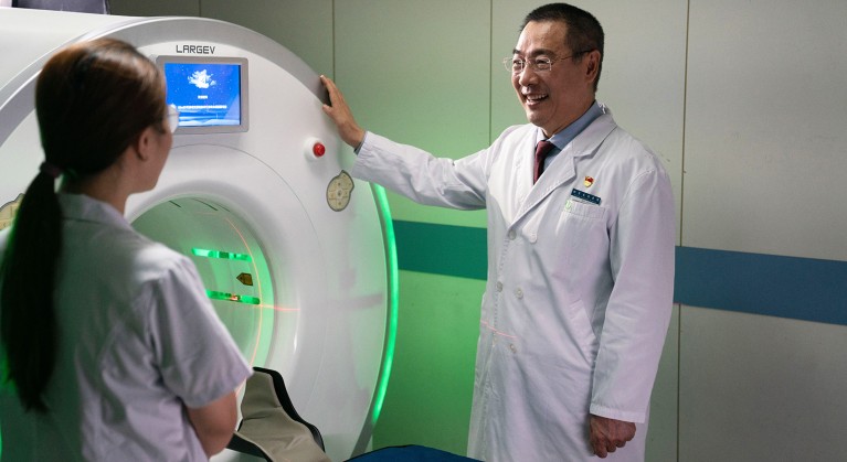
Zhenchang Wang (at right), a radiologist at Beijing Friendship Hospital, leads a team that developed a new ultra-high resolution CT scanner (pictured).
Early diagnosis of some ear diseases is challenging, due to the tiny and intricate nature of a sound-conducting bone called the stapes, made up of four parts. Around 3 millimetres in height, it is one of the tiniest and most complex bones in the human body.
The spatial resolution of standard general-purpose CT (computed tomography) scans is limited to a resolution of 300 micrometres, which is not precise enough to detect small lesions within this bone.
“Resolution is vital for medical imaging,” says Zhenchang Wang, a radiologist with more than 30 years of experience in diagnostic imaging, who is also the vice president of Beijing Friendship Hospital, Capital Medical University in Beijing, China. “Higher resolution scans can more accurately show body parts, especially those with delicate and complex structures.”
Precise imaging
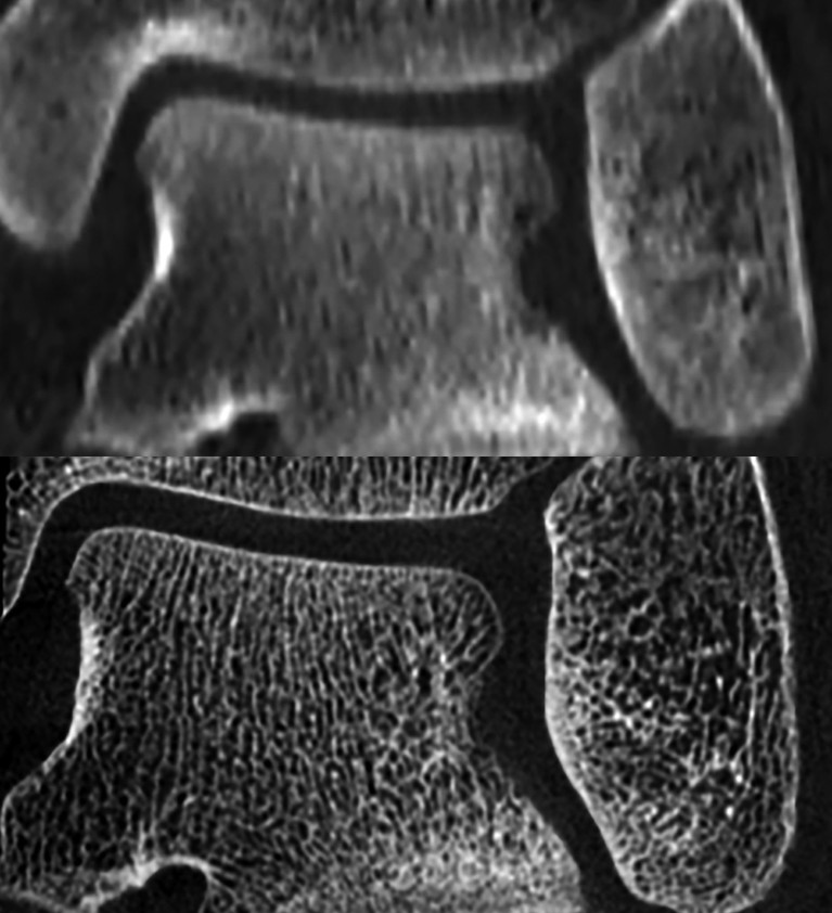
Under Wang’s leadership, the hospital has developed an ultra-high resolution CT technology, specifically for scanning the skeleton, that has a resolution down to 50 micrometres.
“It increases the spatial resolution of the image by a factor of six, and also reduces the radiation dose by two thirds, compared with the previous highest resolution CT,” he says.

The bones of the ankle joint imaged with a conventional CT (top); The ankle bones imaged in more detail with ultra-high resolution CT (bottom).
The technology has also made possible, for the first time, the clear imaging of important anomalies in microstructures that cause hearing disorders1.
Wang says this advance has already improved our understanding of the inner ear labyrinth2, risk factors for facial nerve injuries3, and lesions in the jugular bulb4 — a structure in the skull located close to the ear.
In 2015, Wang was challenged by the low success rate of typical CT scans in diagnosing ear problems. As ear diseases present with few symptoms — including tinnitus, vertigo and hearing impairment — accurate diagnosis relies heavily on visualizing the fine structures of the ear.
However, the resolution of existing CT imaging limited his ability to detect lesions and determine treatment options.
To improve the resolution, Wang worked with a team of physicists, engineers and clinical radiologists from Tsinghua University, Beijing Friendship Hospital, and other renowned hospitals in China to develop a bone-specific CT technology ideal for visualizing bone microstructure.
In 2022, the CT scanning device was approved for use by National Medical Product Administration in China, and the hospital is seeking similar approvals with the relevant bodies in the EU and US.
Eight leading hospitals in Beijing, Shanghai, Guangzhou, and other cities have now used the device to make more than 10,000 CT scans of diseases, such as vertigo, otosclerosis, and temporomandibular joint disorder.
Now, Wang’s team is working to expand applications of the technology. “With ultra-high-resolution imaging, doctors will be able to assess bones throughout the body at a scale down to a scale of 50 microns,” says Wang. Under his lead, orthopedic specialists are exploring its use to shed new light on diseases such as osteoporosis.
Great potential
More specialized imaging equipment is being developed, Wang says.
With support from the National Natural Science Foundation of China, and the Chinese Ministry of Science and Technology, a CT imaging device dedicated to the lungs, and also a new MRI (magnetic resonance imaging) device, are under development.
“Being able to detect abnormalities at an early stage allows for more timely treatment and better outcomes,” he says.


