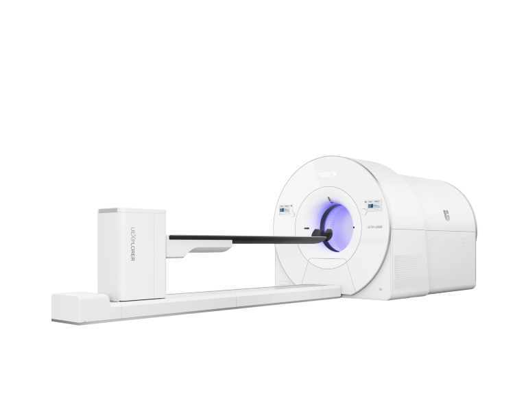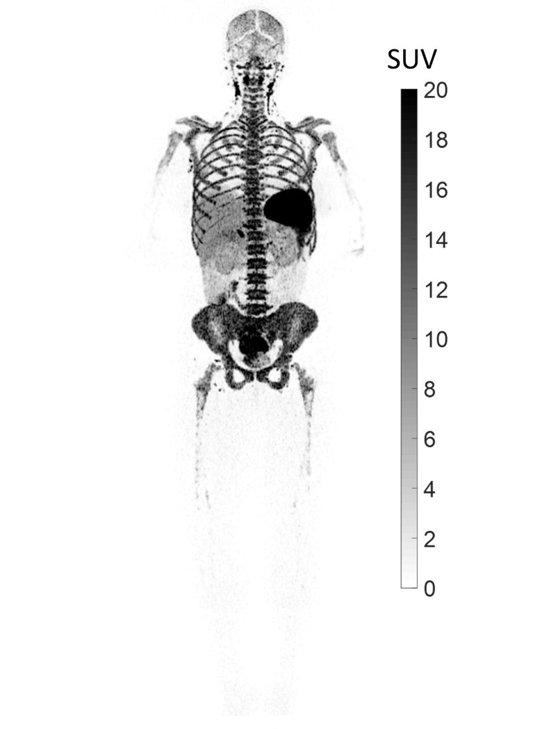
The Total-body PET-CT uEXPLORER.Credit: United Imaging Healthcare
A new medical imaging system is capable of performing full-body scans at high resolution in about half a minute, as opposed to 20–30 minutes for a conventional scan of a part of the body. uEXPLORER, which can also produce dynamic imaging, uses positron-emission tomography (PET) and X-ray computed tomography (CT), and researchers believe it has huge potential to advance medical research.
The technology can shed light on the distribution of T cells produced in response to infectious diseases. Specific T cells known as CD8+ T cells recognize and destroy cells infected by a virus. Despite 95% of CD8+ T cells staying in the body’s tissues rather than in the circulating blood, previous studies have mainly relied on blood samples, which limits knowledge about how many CD8+ T cells are generated, where they accumulate, and their movement in the body.
Now, researchers at the University of California, Davis (UC Davis) are using their Total-body PET–CT scanner to measure the distribution of these immune cells throughout the body1. “It’s the first time we’ve been able to image the distribution of T cells throughout the whole body,” notes Simon Cherry, biomedical engineer, and a co-leader in the total-body scanner development project, along with Ramsey Badawi, who is also vice chair for Research in the Department of Radiology.

Credit: United Imaging Healthcare
The Total-body PET–CT scanner uEXPLORER can image all the tissues and organs of the body in 30 seconds at high sensitivity and high resolution — reducing the imaging time and the radiation dose patients are subjected to, without compromising image quality, which has a resolution of about 2.8 millimeters.
The remarkable device is the fruit of a collaboration between UC Davis and United Imaging Healthcare, an innovator in advanced medical imaging based in Shanghai, China, that has headquarters and a factory in Texas, in the United States.
A disruptive technology
Conventional PET–CT scanners are used to detect conditions including cancers, heart disease and brain disorders. Patients are injected with a radioactive tracer and after about an hour, the patient slowly moves through a ring-shaped detector, which detects radiation from the tracer. One scan takes about 20 minutes, and the scanner then constructs a 3D map of the cells metabolizing the tracer molecule.
A conventional detector is just 20 centimeters across, and so to cover a large area, the patient needs to move through the scanner, allowing it to pass over the whole body.
United Imaging Healthcare’s new device is nearly 2 meters long and consists of eight detector rings that can simultaneously scan an adult body. This allows it to collect radiation emitted in all directions, with 40 times the sensitivity of conventional PET2.
“It captures radiation much more efficiently, which means we get a lot more signal and can thus image very quickly,” says Cherry.
A scan time of 30 seconds can achieve an equivalent diagnostic quality as conventional PET–CT scanners in clinical practice, according to a study by Hongcheng Shi, head of nuclear medicine at Zhongshan Hospital in Shanghai, China.
A long journey
The goal of imaging the whole human body in a single scan goes back 17 years to when Cherry, who was developing PET scanners for imaging small animals for pharmaceutical research, met Badawi, who was running computer simulations to realize larger field of views for human scanners.
“At that time, we were already taking the full image of small model animals like mice,” recalls Cherry. “We thought why don’t we just scan the whole human body? The benefits could be boundless.”
It took them a decade to obtain funding for the bold project; eventually the National Institutes of Health supported it in 2015. The next challenge was finding the right industry partner to commercialize it. “The complexity of this system rivals that of some of the high-energy physics experiments at CERN [European Organization for Nuclear Research],” explains Cherry.

Simon Cherry (right) and Ramsey Badawi (seated), professors from University of California, Davis, with Hongdi Li (left) and the team from United Imaging Healthcare.Credit: United Imaging Healthcare
The research caught the attention of Hongdi Li, chief technical officer of United Imaging Healthcare in China and chief executive of United Imaging Healthcare America. "Our team likes to innovate, and our technology is sufficient for such challenges," says Li.
One obstacle for such a complex system is collecting signals. Li’s team proposed using silicon photomultipliers to digitize the raw signal for a compact modular design, allowing for higher sensitivity and image resolution. "United Imaging Healthcare had already applied digital PET technology based on silicon photomultipliers to our products and achieved high-resolution imaging," notes Li. The journey then entered a new phase.
Broad potential
In 2019, the scanner was approved by the US Food and Drug Administration. It was then installed at the EXPLORER Molecular Imaging Center in UC Davis and at Zhongshan Hospital, Shanghai, China.
Increasingly, research teams are exploring the total-body scanner’s exciting potential. Its ability to perform dynamic imaging allows near real-time observation of drug distribution in the body. Shi’s team uses three different doses of radiation tracer — full dose, half dose and a tenth of a dose — based on the patient’s vulnerability to radiation. The scanner can both help enhance clinical services and pre-clinical research, Shi says.
For example, previously scientists could image only where the tracer accumulated in the body. They can now observe the tracer distribution throughout the body in near real time, allowing them to determine the biological distribution of drugs and glean visual evidence for the future integration of diagnosis and treatment, Shi says.
The scanner can be used to study chronic systemic diseases such as autoimmune inflammatory arthritis, which causes joint inflammation. Scientists can map the joints and visualize inflammation with an extremely high spatial resolution3.
Many more projects using the scanner are underway. Biotechnology company BAMF Health in the US is using the scanner to provide precision treatment for cancer patients; Renji Hospital in China is employing it to study the brain-gut axis; UC Davis and the University of California, San Francisco, are using new tracers to map the distribution of HIV viral load in the body.
The journey continues. United Imaging Healthcare and UC Davis are continuing to collaborate, this time on next-generation brain-scanning projects, along with Yale University. United Imaging Healthcare plans to build a community for medical engineering, aiming to encourage PET users to share their latest discoveries and tools. “We look forward to more collaborations with academic institutions, to find tailored, precise solutions and bring the value of molecular imaging for high-quality patient care,” says Li.
This advertisement appears in Nature Index 2022 Innovation, an editorially independent supplement. Advertisers have no influence over the content of the editorial articles within this supplement.


