A new scanning strategy makes it possible to record network physiology from viewing angles previously inaccessible, revealing 3D processing characteristics of the intact neural net. Credit: Máté Marosi & Csaba Csupernyák, Femtonics
Botond Roska investigates the computations that brain circuits execute on a moment-by-moment basis. His approach is conceptually straightforward — but technically demanding. Experiments in his lab at the Institute of Molecular and Clinical Ophthalmology Basel in Switzerland involve recording the activity of single cortical neurons — plus the activity of up to 200 neighbouring neurons that provide the cell with synaptic inputs.
His group scans these neurons using two-photon (2P) imaging. However, conventional 2P imaging systems operate only in a 2D plane, but neuronal networks are dispersed in three-dimensional space. The problem lies in the way most 2P systems focus the laser, scanning continuously along lines or planes. Moving beyond a plane typically requires mechanical movement of the lens, which slows down sampling rates considerably.
When Roska’s team scanned the 3D network plane-by-plane using 2P imaging, they had to sequentially characterize all the presynaptic neurons’ activity, then infer how these qualities could yield the properties of the target neuron. When they published the results in 2015, they included a supplementary figure showing how an alternative type of imaging could be used to do these experiments1.
This other technology, called random-access 3D microscopy, allows a 2P laser to move almost instantaneously between many points within a region. “What you can now do”, Roska says, “is look simultaneously at all 150-200 presynaptic neurons that give input to your postsynaptic neuron and what they do at a given moment. You can really start to analyse how the brain computes in real time.”
Sound and vision
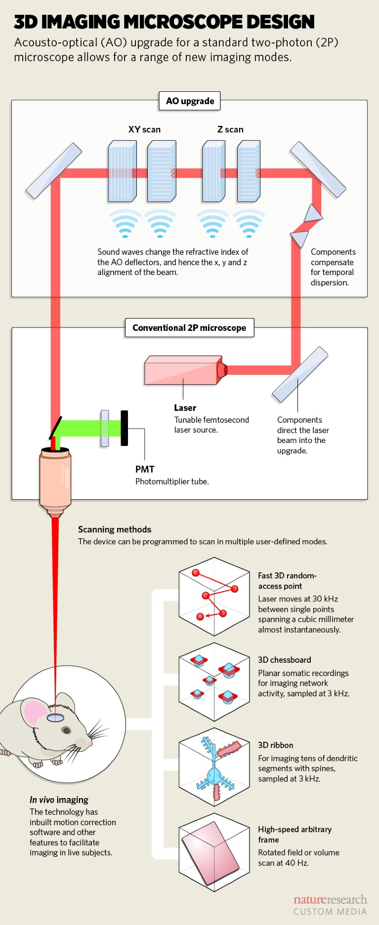
This 3D scanning is achieved by inserting an upgrade containing acousto-optical (AO) deflectors in the laser path of a 2P microscope. An AO deflector is essentially a transparent crystal with a refractive index that can be modulated by passing soundwaves through it. Changing the frequency of those soundwaves changes where the laser focuses, allowing it to be directed anywhere in a 3D space in microseconds (see ‘3D imaging microscope design’).

Ivo Vanzetta, a neuroscientist at the Institut de Neurosciences de la Timone, in Marseille, France, has also used random-access 3D microscopy to image neuronal calcium signals – not just from hundreds of neurons, but from over one thousand2.
“In a chunk of cortex,” Vanzetta says, “what you’re interested in is the cell bodies and dendrites, and these are distributed sparsely. Most of the volume is not of interest.” With random access 3D microscopy, the experimenter programs the points of interest, meaning data are only collected from what’s important. This both speeds up sampling rates and minimizes photodamage to the tissue.
Both Vanzetta and Roska use 3D systems developed by Femtonics, a company founded in 2005 by researchers from the Hungarian Academy of Sciences in Budapest. They are one of three academic groups3,4 that independently developed AO devices for 2P imaging, but the only one making the technology commercially.
The Budapest group were also the first to publish methods5 for high-speed recording of neuronal activity in vivo in 2012. Then, in 2016, improved scanning technology and motion correction algorithms enabled imaging in awake, behaving mice. Not only could it image cell bodies across a large volume of brain, but it could also observe activity in hundreds of spines across the complex dendritic arbour of individual neurons6.
The company has worked with several academic collaborators to identify researchers’ needs, says Gergely Szalay, Femtonics’ research and development manager and a research fellow at the Hungarian Academy of Sciences. The latest system is the Femto3D Atlas with improved AODs to enable 3D scanning as well as a fast plane-scanning mode with the freedom of arbitrary rotation in space. “We can jump anywhere in three dimensions and also emulate all the traditional scanning modes,” says Szalay. “This unites the benefits of galvanometric and resonant scanner-based microscopes with 3D random-access scanning.”
The Femto3D Atlas can be a standalone multiphoton microscope or an upgrade to a 2P microscope. It samples fast, has an excellent signal-to-noise ratio, and motion-correction software for in vivo work.

Femtonics’ research and development manager, Gergely Szalay, and applications specialist Tamás Tompa (both seated) show the Femto3D Atlas to interested researchers.Credit: Femtonics
Roska is excited; over the course of nearly a decade, 3D imaging has grown from an interesting idea to the workhorse microscopes of his lab. “Right now,” he says, “it’s a really mature technology. The science over the next few years will show just what we can pull out.”


