Researchers at City University of Hong Kong (CityU) have made significant advances in unravelling fundamental mechanisms behind biological processes and diseases, especially in neuroscience, cancer biology, and regenerative medicine. Their work opens new avenues for developing diagnostic and therapeutic strategies.
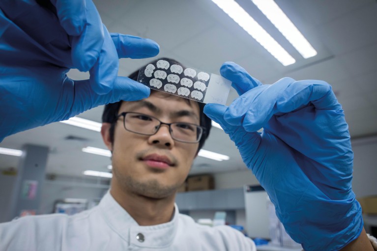
CityU biomedical scientists have shed light on how memory is formed by clarifying the mechanism of cholecystokinin (CCK), which is also a digestive hormone.
CityU’s latest research adds to knowledge about memory formation mechanisms. Credit: City University of Hong Kong
New understanding to memory formation mechanism
The function of CCK as the brain’s memory-writing switch was raised a few years ago by a team led by He Jufang, Chair Professor of Neuroscience at CityU. Recently, in work published in the Proceedings of the National Academy of Sciences of the United States of America (PNAS), the team further revealed that high-frequency stimulation (HFS) induces the release of CCK, which is controlled by N-methyl-D-aspartate (NMDA) receptors, leading to memory formation.
Memory is stored in neural networks through changes in synaptic strength. Long-term potentiation (LTP), a long-lasting strengthening of synapses, is believed to represent the neural basis of memory. HFS-induced neocortical LTP has largely been thought to be dependent on NMDA receptors. He’s team demonstrated, however, that the LTP formation process is CCK-dependent. As NMDA receptors are critical in triggering the release of CCK, by infusing CCK into the cortex, LTP can be induced even after blocking NMDARs receptors. The work consolidates the theoretical basis for developing new treatments for Alzheimer’s disease and epilepsy.
Enabling gene therapies against cancer
While gene therapies offer great potential for cancer treatment, a safe and effective drug delivery strategy remains elusive. The difficulty is that common vehicles for delivering RNA drugs, including viruses, lipid transfection reagents and lipid nanoparticles, are usually immunogenic and/or cytotoxic. A research project, led by Le Minh and Shi Jiahai, from CityU’s Department of Biomedical Sciences (BMS) sought new drug carriers.
Extracellular vesicles (EVs), like natural cellular couriers, facilitate intercellular communication by transporting proteins and other large molecules through the membrane between cells, and can be good candidates for drug carriers. However, the stringent purification methods currently used to produce EVs are time-consuming and demand billions of cells, so are not scalable.
The innovation by the CityU team, published in Nature Communications, involves producing EVs from human red blood cells, which are abundant, and lack nuclear or mitochondrial DNA. Their strategy could generate 1,000 times more EVs at a 100th of the cost, compared to the existing method. Using these EVs, RNA drugs could be delivered to 80% of the cancer cells, a significant improvement in efficiency, and showed no side-effects.
Safer gene therapy vectors to treat retinal degeneration diseases
Approved by the US Food and Drug Administration (FDA), adeno-associated virus (AAV) vector is proven for safety and efficiency. The AAV-based gene therapy has brought great hope to people with inherited blindness.

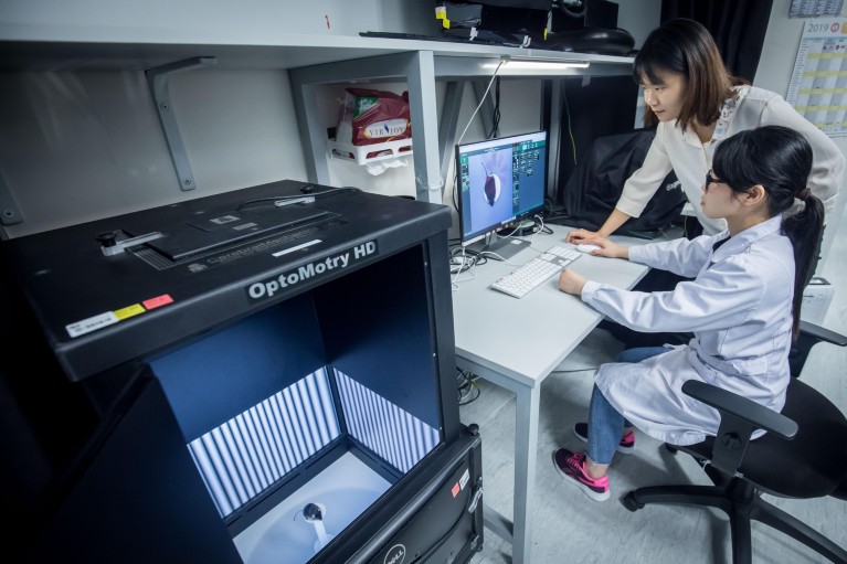
Dr. Xiong Wenjun (right) found some adeno-associated viral vectors used in gene therapy could cause inflammation and toxicity in the eyes of mice. Credit: City University of Hong Kong
However, collaborating with colleagues from Harvard Medical School, Xiong Wenjun, a BMS assistant professor, recently discovered that some AAV vectors could cause inflammation and toxicity in the eyes of mice, inducing retinal cell death and severe visual function loss. The team further found that the toxicity was associated with certain cis-regulatory sequences which control gene expression in the virus genome. The toxic cis-regulatory sequences commonly drive broad and strong gene expression in the eyes, especially in retinal pigment epithelial cells. Published in PNAS, the study reveals that if sensitive and organ-specific assays are developed and tested for different manifestation of toxicity, safer vectors could be designed to avoid inflammation and toxicity due to viral vector genomic elements.
A biomarker for categorizing colorectal cancer patients
Colorectal cancer (CRC) is one of the leading causes of cancer-related deaths worldwide. While endoscopic submucosal dissection (ESD), a minimally invasive surgical procedure to remove gastrointestinal tumours, is sufficient for treating early-stage CRC patients who are at low risk for developing lymph-node metastasis (LNM), the current risk-stratification criteria based on post-endoscopic pathological examination tend to overestimate the degree of risk. Many supposedly low-risk patients might be inadvertently categorized as high-risk, and undergo unnecessary surgeries.
A study by Wang Xin, a BMS assistant professor, in collaboration with colleagues from Baylor University Medical Center, and the University of Tokyo, recently reported in Gastroenterology the development of a novel 8-gene mRNA classifier for more accurate detection of LNM in patients with T1 CRC. In their comparison study, current pathological criteria for risk assessments would stratify 84% patients in the high-risk category, of whom only 17% were found LNM-positive in post-surgical examination of tissues; the novel mRNA classifier would only categorize 23% of patients as high-risk, of whom 48% were actually LNM-positive. The new approach can reduce unnecessary treatment T1 CRC patients by nearly 60%.
Collecting respiratory droplets on bio-inspired surfaces
Airborne fluids spread by coughs and sneezes are key to the spread of disease. Surface tension allows these pathogen-bearing micro-droplets to adhere to surfaces and cause contamination. Exerting external electrical force could help repel micro-droplets from point-of-care biomedical devices, but an optimum design has been elusive so far.
Inspired by naturally occurring biological processes, such as the ability of some insects to use surface tension to walk on water, Yao Xi, a BMS assistant professor, and his team developed an innovative method for moving and collecting micro-droplets without energy input. Observing the way in which seeds float and aggregate on water surface in water gardens, they printed anti-bacterial hydrogel dots on a liquid-infused surface, creating an asymmetric curve. With the help of capillary forces, the micro-droplets are pumped towards the hydrogel dots continuously and efficiently, and bacteria are detected and destroyed. Their study was published in PNAS, demonstrating an energy-efficient technique for bacteria detection and disinfection.
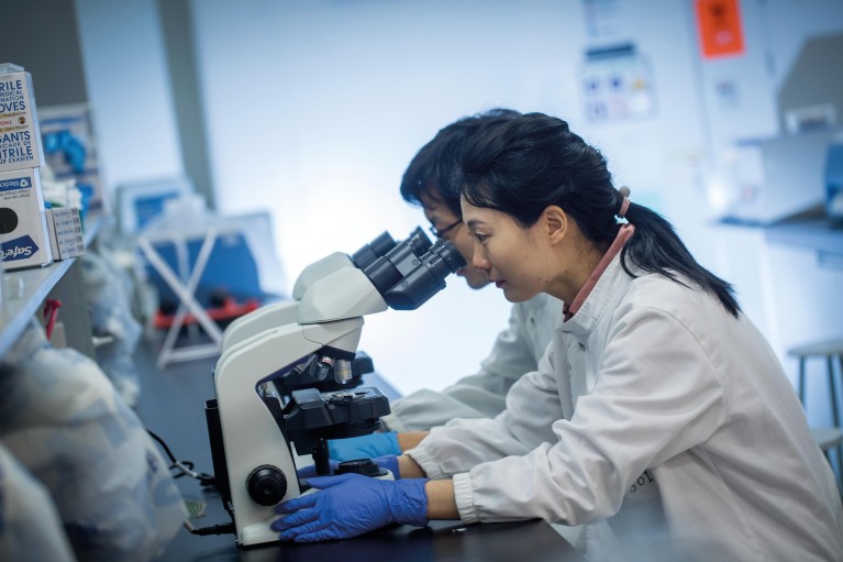
Moving forward, the critical mass of biomedical scientists at CityU will continue working towards a common goal: translating their discoveries into practical applications for the benefit of human health.
Biomedical scientists at CityU will continue working hard to translate discoveries into practical applications for the benefit of human health. Credit: City University of Hong Kong


