James Watson and Francis Crick are two of the twentieth century’s most renowned scientists. The seminal paper from the pair at the University of Cambridge, UK, detailing the discovery of the DNA double helix, was published as part of a trio in Nature 70 years ago this week1–3. They are also widely believed to have hit on the structure only after stealing data from Rosalind Franklin, a physical chemist working at King’s College London.
Lore has it that the decisive insight for the double helix came when Watson was shown an X-ray image of DNA taken by Franklin — without her permission or knowledge. Known as Photograph 51, this image is treated as the philosopher’s stone of molecular biology, the key to the ‘secret of life’ (not to mention a Nobel prize). In this telling, Franklin, who died of ovarian cancer in 1958 at just 37, is portrayed as a brilliant scientist, but one who was ultimately unable to decipher what her own data were telling her about DNA. She supposedly sat on the image for months without realizing its significance, only for Watson to understand it at a glance.
This version of events has entered into popular culture. It is the subject of Photograph 51, a play by Anna Ziegler that starred Nicole Kidman on the London stage in 2015. The image graces a British 50 pence coin that marked the centenary of Franklin’s birth, in 2020. The whole affair has provided fodder for scornful Twitter jokes (“What did Watson and Crick discover in 1953? Franklin’s data.”) and even a marvellous rap battle by seventh-grade students in Oakland, California.
But this is not what happened.
How Rosalind Franklin was let down by DNA’s dysfunctional team
One of us (N.C.) is writing a biography of Watson, the other (M.C.) is writing one of Crick. In 2022, we visited Franklin’s archive at Churchill College in Cambridge, UK, and went through her notes together, reconstructing the development of her ideas. We also found a hitherto unstudied draft news article from 1953, written in consultation with Franklin and meant for Time, a US magazine with international reach — as well as an overlooked letter from one of Franklin’s colleagues to Crick. Together, these documents suggest a different account of the discovery of the double helix. Franklin did not fail to grasp the structure of DNA. She was an equal contributor to solving it.
Getting Franklin’s story right is crucial, because she has become a role model for women going into science. She was up against not just the routine sexism of the day, but also more subtle forms embedded in science — some of which are still present today.
Franklin and DNA
In the early 1950s, the structure and function of DNA remained unclear. It had been found in every cell type investigated, and was known to consist of a phosphate backbone to which were attached four kinds of base — adenine, thymine, cytosine and guanine (A, T, C and G).
In 1944, the microbiologist Oswald Avery and his colleagues had shown that DNA (not protein) could transform benign Streptococcus pneumoniae bacteria into a virulent form4. But it remained far from clear that it was the genetic material in all organisms.
At King’s College London, biophysicists funded by the Medical Research Council (MRC), and led by John Randall, with Maurice Wilkins as his deputy (who would later share the Nobel prize with Watson and Crick in 1962), were using X-ray diffraction to study the structure of the molecule. In 1951, they were joined by Franklin, who had been using this technique to investigate the structure of coal at the Central State Laboratory of Chemical Services in Paris.

Maurice Wilkins (left), James Watson and Francis Crick at the ceremony for the 1962 Nobel Prize in Physiology or Medicine.Credit: King’s College London Archives: K/PP178/15/3/1
As is well known, Franklin and Wilkins clashed, in both personality and scientific approach. Although Franklin relished a good argument and was determined to make progress, Wilkins abhorred confrontation and was slower to act. To ease tensions, Randall divvied up the DNA work. In what Wilkins later called a bad bargain for himself, he agreed to turn over to Franklin the small supply of very pure DNA that he had obtained from the Swiss chemist Rudolf Signer. Wilkins was stuck with poorer quality stuff from the Austrian biochemist Erwin Chargaff, at Columbia University in New York City.
With the Signer DNA, Franklin was able to exploit a discovery that Wilkins had made earlier — DNA in solution could take two forms, what she called the crystalline or A form, and the paracrystalline or B form. Franklin found that she could convert A into B simply by raising the relative humidity in the specimen chamber; lowering it again restored the crystalline A form.
Franklin focused on the A form, Wilkins on the B form. To a physical chemist, the crystalline form seemed the obvious choice. When bombarded with X-rays in front of a photographic plate, it yielded sharp, detailed diffraction patterns. More detail meant more data, which meant a more accurate, albeit more difficult analysis. The B form, by contrast, yielded patterns that were blurrier and less detailed, but simpler to analyse. Initially, Franklin understood both A and B as helical. In notes for a seminar she gave in November 1951, she described them collectively: “big helix with several chains, phosphates on outside, phosphate–phosphate interhelical bonds, disrupted by water”5.
Unable to resolve the A-form structure, Franklin had decided by the middle of 1952 that it was not actually helical — she even teased Wilkins with a mock funeral notice for the crystalline DNA helix6. She was not alone in being thrown off by the A-form data: after the double-helix paper1 had been published, Crick wrote of Franklin’s precise but complex, data-rich A-form image, “I am glad I didn’t see it earlier, as it would have worried me considerably”7.
The double helix and the ‘wronged heroine’
As for the B form, she and everyone else at King’s recognized that it was some kind of helix. But to Franklin it was a distraction. At high humidity, water molecules crowded the atoms in DNA, producing a structure she described as “swollen”, “distended”, disordered. “Anyway,” she wrote in the notes for her 1951 seminar, under increased humidity, “the stuff ultimately dissolves, i.e. chains are separated from one another by water”5. She saw the B form as an artefact of being water-logged, a symptom of the loss of crystalline order — hence “paracrystalline”. This explains why, in late 1952 and early 1953, she rejected the argument that DNA was intrinsically helical.
From a chemist’s perspective, Franklin’s decision to focus on the crystalline A form was perfectly logical, as were the conclusions she drew from analysing it. But her focus on the drier A form ignored the very wet reality of the inside of a cell — which would mean that DNA took the more humid B form. Together with her insistence that the diffraction data be fully analysed before any modelling was attempted, it would hamper Franklin’s efforts for more than a year.
The meaning of Photograph 51
Even Franklin’s advocates often unwittingly perpetuate a caricatured view of her science — one that can be traced back to Watson’s reality-distorting 1968 bestseller, The Double Helix8. Watson’s version of the next, crucial stage in the story is often repeated to highlight how Franklin was deprived of due credit. Inadvertently, this undermines her.
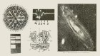
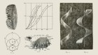
According to Watson, in early 1953, he visited King’s and got into a row with Franklin. Wilkins, he wrote, rescued him from the confrontation and then showed him Photograph 51, a particularly clear image of the B form, taken 8 months earlier by Franklin and her graduate student Raymond Gosling. Franklin had put the photograph aside to concentrate on the A form. She was preparing to transfer to Birkbeck College, also in London, and had been instructed to leave her DNA work behind. Gosling was now being supervised by Wilkins, and he had given Wilkins the photograph. (He says he did so with Franklin’s knowledge9.) The image, Watson claimed in The Double Helix, showed that a DNA helix “must exist” — only a helical structure could produce those marks8.
Because of Watson’s narrative, people have made a fetish of Photograph 51. It has become the emblem of both Franklin’s achievement and her mistreatment.

Franklin and Gosling’s X-ray diffraction image of B DNA, known as Photograph 51.Credit: King's College London Archives/Science Photo Library
But Watson’s narrative contains an absurd presumption. It implies that Franklin, the skilled chemist, could not understand her own data, whereas he, a crystallographic novice, apprehended it immediately. Moreover, everyone, even Watson, knew it was impossible to deduce any precise structure from a single photograph — other structures could have produced the same diffraction pattern. Without careful measurements — which Watson has insisted he did not make — all the image revealed was that the B form was probably some kind of helix, which no one doubted. Furthermore, various lines of evidence — including The Double Helix itself, read carefully — show that it played little, if any, part in Watson and Crick’s inching towards the correct structure between January and March 1953. In fact, it was other data from Franklin and Wilkins that proved crucial, and even then, what really happened was less malicious than is widely assumed.
Watson did get a jolt from seeing the photograph — because of when he saw it. Just days before, the Cambridge group had received a manuscript from the US chemist Linus Pauling, in which he’d claimed to have solved the DNA structure. Although Pauling had made some elementary errors, Lawrence Bragg, head of the Cavendish Laboratory, who had a long-standing rivalry with Pauling, had encouraged Watson and Crick to resume their model building. Watson had dropped in at King’s to show off Pauling’s blunder, and Wilkins had shown him the photograph. Fashioning that moment into the climax of The Double Helix was a literary device: a classic eureka moment, easy for lay readers to understand.
From 1951, Wilkins had kept Watson and Crick abreast of his work on the B form, in particular his belief that the structure contained one or more helices, repeated every 34 angstroms, and he might have said that within each repeat there were probably 10 elements. Shortly after Watson saw Photograph 51, Crick’s supervisor, Max Perutz, handed them an informal report of the activity of the King’s MRC unit, which he had been given as part of an official visit to the unit in December 1952. This included a page from Franklin, describing her work. In a 1969 letter to Science, although Perutz said that he regretted sharing the report without first consulting the King’s group, it was not confidential10. Indeed, a letter we have discovered from a King’s researcher, Pauline Cowan, written to Crick in January 1953, invites Crick to a talk by Franklin and Gosling, who, Cowan continues, “say that it is mostly for a non-crystallographic audience + that Perutz already knows more about it than they are likely to get across so you may not think it worthwhile coming”. Thus, Franklin seems to have assumed that Perutz would share his knowledge with Crick as part of the usual informal scientific exchange11.
In her contribution to the MRC report, Franklin had confirmed the 34 Å result for the B form. She also reported that the unit cell (the repeating unit of the crystal) of DNA was huge; it contained a larger number of atoms than any other unit cell in any other known molecular structure. Franklin also added some key crystallographic data for the A form, indicating that it had a ‘C2’ symmetry, which in turn implied that the molecule had an even number of sugar-phosphate strands running in opposite directions.
Notes by Crick for a lecture on the history of the double helix, given to historians of science at the University of Oxford in May 1961, together with formal and informal remarks made throughout his life, reveal that, unlike Photograph 51, this report was truly significant for confirming the structure that Watson and Crick eventually obtained.
In the end, however, neither Photograph 51 nor the MRC report ‘gave’ Watson and Crick the double helix. What did was six weeks of what they later described as “trial and error” — making chemical calculations and fiddling about with cardboard models. (Watson made this plain in The Double Helix; Crick did so in a series of interviews with the historian Robert Olby in the late 1960s and early 1970s.)
Franklin’s data and Watson and Crick’s many conversations with Wilkins had provided what seem like key pieces of information — the phosphate groups were on the outside of the molecule; there was a repeat every 34 Å; perhaps there were ten bases per repeat and an even number of strands running in opposite directions (the implication of the C2 symmetry). Yet, according to their own accounts, the pair ignored every one of these facts at one point or another during those six weeks. Once they had hit on a conceptual model of the structure, the MRC report provided a valuable check on their assumptions.
So it was not a case of them stealing the King’s group’s data and then, voila, those data gave them the structure of DNA. Instead, they solved the structure through their own iterative approach and then used the King’s data — without permission — to confirm it.
What Franklin really did
Franklin contributed several key insights to the discovery of the double helix. She clearly differentiated the A and B forms, solving a problem that had confused previous researchers. (X-ray diffraction experiments in the 1930s had inadvertently used a mixture of the A and B forms of DNA, yielding muddy patterns that were impossible to fully resolve.) Her measurements told her that the DNA unit cell was enormous; she also determined the C2 symmetry exhibited by that unit cell12.
The C2 symmetry was one of 230 types of crystallographic 3D ‘space groups’ that had been established by the end of the nineteenth century. Franklin failed to appreciate its significance not because she was obtuse, but because she was unfamiliar with it. According to her colleague Aaron Klug, Franklin later said that she “could have kicked herself” for not realizing the structural implications13. Crick did realize the implications because he happened to have studied C2 symmetry intensely. But even he did not use Franklin’s determination of this symmetry when building the model; rather, it provided a powerful corroboration when their model was complete.

James Watson (left) and Francis Crick modelled the structure of the DNA double helix.Credit: A. Barrington Brown, Gonville & Caius College/Science Photo Library
Franklin also grasped, independently, one of the fundamental insights of the structure: how, in principle, DNA could specify proteins. In February 1953, she was working hard to finish her analyses of DNA before leaving King’s. The A form had continued to resist her attempts to interpret it, so she had turned to the much simpler, clearly helical B form. Her notes reveal that by late February, she had accepted that the A form was also probably helical, with two strands, and she had realized that the order of the bases on a given strand had no effect on the overall structure. This meant that any sequence of bases was possible. As she noted, “an infinite variety of nucleotide sequences would be possible to explain the biological specificity of DNA”14. This idea, which Watson and Crick grasped at around the same time, had first been proposed in 1947 by chemist John Masson Gulland at University College Nottingham, UK (now the University of Nottingham)15.
Franklin did not apprehend complementary base-pairing — that A could bond only with T and C only with G, with each pair of bases forming an identical structure in the molecule. In fact, she was not working with the correct forms of the bases, so she could not have made a satisfactory model had she tried (the same was true of Watson and Crick until the very last phase of their work). Neither did she realize that her data implied that the two strands were oriented in different directions — or that the B form, found at high levels of humidity, must be the biologically functional form. (The A form is found only under laboratory conditions.) She did not have time to make these final leaps, because Watson and Crick beat her to the answer.
Franklin did not succeed, partly because she was working on her own without a peer with whom to swap ideas. She was also excluded from the world of informal exchanges in which Watson and Crick were immersed. Even though some at the time — notably the researchers at King’s and a small flock of what Watson called “minor Cambridge biochemists”16 — were not happy about Watson and Crick’s use of the King’s group’s data, the lead scientists at the Cavendish — Perutz, Bragg, John Kendrew — thought it was quite normal. And there is no evidence that Franklin thought otherwise.
Acknowledging the truth
After Watson and Crick had read the MRC report, they could not unsee it. But they could have — and should have — requested permission to use the data and made clear exactly what they had done, first to Franklin and Wilkins, and then to the rest of the world, in their publications.
In April 1953, Nature published three back-to-back papers on DNA structure, from Watson and Crick, from Wilkins and his co-workers, and from Franklin and Gosling1–3. Watson and Crick declared that they had been “stimulated by a knowledge of the general nature of the unpublished experimental results and ideas” of Wilkins and Franklin. They insisted, though, that they were “not aware of the details”, claiming that the structure “rests mainly though not entirely on published experimental data and stereochemical arguments”1. The truth of those statements depends on highly charitable interpretations of “details” and “mainly though not entirely”.
Rosalind Franklin was so much more than the ‘wronged heroine’ of DNA
In a full description of the structure in a paper submitted in August 1953 and published in 1954, Crick and Watson did attempt to set the record straight17. They acknowledged that, without Franklin’s data, “the formulation of our structure would have been most unlikely, if not impossible”, and implicitly referred to the MRC report as a “preliminary report” in which Franklin and Wilkins had “independently suggested that the basic structure of the paracrystalline [B] form is helical and contains two intertwined chains”. They also noted that the King’s researchers “suggest that the sugar-phosphate backbone forms the outside of the helix and that each chain repeats itself after one revolution in 34 Å”.
This clear acknowledgement of both the nature and the source of the information Watson and Crick had used has been overlooked in previous accounts of the discovery of the structure of DNA. As well as showing the Cambridge duo finally trying to do the right thing, it strengthens our case that Franklin was an equal member in a group of four scientists working on the structure of DNA. She was recognized by her colleagues as such, although that acknowledgement was both belated and understated. All this helps to explain one of the lasting enigmas of the affair — why neither Franklin nor Wilkins ever questioned how the structure had been discovered. They knew the answer, because they expected that Perutz would share his knowledge and because they had read Watson and Crick’s 1954 article17.
Time out
Three weeks after the three DNA papers were published in Nature, Bragg gave a lecture on the discovery at Guy’s Hospital Medical School in London, which was reported on the front page of the British News Chronicle daily newspaper. This drew the attention of Joan Bruce, a London journalist working for Time. Although Bruce’s article has never been published — or described by historians, until now — it is notable for its novel take on the discovery of the double helix.
Bruce portrayed the work as being done by “two teams”: one, consisting of Wilkins and Franklin, gathering experimental evidence using X-ray analysis; “the other” comprising Watson and Crick, working on theory. To a certain extent, wrote Bruce, the teams worked independently, although “they linked up, confirming each other’s work from time to time, or wrestling over a common problem”. For example, Watson and Crick had “started to work on the double helix theory as a result of Wilkins’ X-rays”. Conversely, she wrote, Franklin was “checking the Cavendish model against her own X-rays, not always confirming the Cavendish structural theory”18. It has not escaped our notice that both examples render Franklin in a position of strength, every bit a peer of Wilkins, Crick and Watson.
Unfortunately, Bruce was not so strong on the science. Her article got far enough for Time to send a Cambridge photographer, Anthony Barrington Brown, to shoot portraits of Watson and Crick, and for Watson to tell his friends to watch for it19. But it never appeared, perhaps because Franklin told Bruce that it needed an awful lot of work to get the science straight. Bruce’s take on the discovery was buried, and Barrington Brown’s compelling images disappeared until Watson resurrected the best of them 15 years later, for The Double Helix20.
It is tantalizing to think how people might remember the double-helix story had Bruce’s article been published, suitably scientifically corrected. From the outset, Franklin would have been represented as an equal member of a quartet who solved the double helix, one half of the team that articulated the scientific question, took important early steps towards a solution, provided crucial data and verified the result. Indeed, one of the first public displays of the double helix, at the Royal Society Conversazione in June 1953, was signed by the authors of all three Nature papers1–3,21. In this early incarnation, the discovery of the structure of DNA was not seen as a race won by Watson and Crick, but as the outcome of a joint effort.
According to journalist Horace Freeland Judson and Franklin’s biographer, Brenda Maddox, Rosalind Franklin has been reduced to the “wronged heroine” of the double helix22,23. She deserves to be remembered not as the victim of the double helix, but as an equal contributor to the solution of the structure.
 How Rosalind Franklin was let down by DNA’s dysfunctional team
 The double helix and the ‘wronged heroine’
 Rosalind Franklin was so much more than the ‘wronged heroine’ of DNA