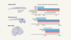
Healthy heart circulation
Deoxygenated blood (blue) arrives in the heart’s right atrium from two veins — the superior and inferior vena cava and is then passed through the tricuspid valve to the right ventricle, which pumps it to the lungs. Oxygenated blood (red) from the lungs travels through the pulmonary veins to the heart’s left atrium, then through the mitral valve to the left ventricle, which pumps the blood out to the rest of the body.

Infographic: Alisdair Macdonald
Three kinds of defect
The anatomy of each SVD differs, but they all result in a mix of oxygenated and deoxygenated blood being pumped to the body and lungs. This means that vital organs don’t get enough oxygen.

Infographic: Alisdair Macdonald
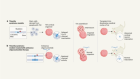
A solution in stages
SVDs are treated with a sequence of three operations2. Together, these surgeries ensure that the deoxygenated blood returning from the body bypasses the heart and goes straight to the lungs. The heart is then left to pump only oxygenated blood — getting rid of the problem of mixed blood.

Infographic: Alisdair Macdonald
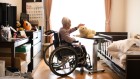
Comorbidities are common
Complications: Children born with SVDs can live into their 30s and 40s, but they have a significantly higher chance of experiencing other diseases as compared with healthier adults3–7.

Infographic: Alisdair Macdonald
In utero surgery
Surgeons have begun to pioneer procedures to start treatment from within the womb.

Infographic: Alisdair Macdonald
 The surgical solution to congenital heart defects
 Video: Babies with misshapen hearts