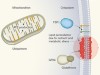
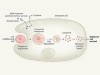
Ghostly metabolic messages from dying cells
“Marley was dead, to begin with. There is no doubt whatever about that.” The iconic opening lines of Charles Dickens’s novel A Christmas Carol convey the idea of the finality of death, a concept that pervades our thinking even when considering the demise of cells. A dead cell is, to echo Dickens’s description of Marley, “as dead as a doornail”. But just as Marley had an influence from beyond the grave to change the character of Ebenezer Scrooge in the story, cells that die can have a vital effect on the living cells around them. Writing in Nature, Medina et al.1 bring this process to life by uncovering metabolic processes in dying cells that have important consequences for the organism.
Every second, millions of cells die in our bodies owing to processes that are a normal part of life, such as tissue turnover and responses to environmental stresses2. The vast majority of these deaths occur by a process called apoptosis. This is a form of cellular suicide that is orchestrated by the actions of enzymes called caspases, which cleave hundreds of different intracellular proteins3. This regulated cleavage of various caspase targets effectively ‘packages’ the dying cell through an orderly dismantling process. DNA in the nucleus is cut into small pieces, the cytoplasmic ‘skeleton’ of filamentous actin protein is remodelled to break the cell into smaller fragments, and the exposure of a particular lipid on the cell surface signals to immune cells, such as macrophages, to take up (engulf) and digest the dying cell2.
Ever since the original description of apoptosis4, it has been known that this form of cell death does not trigger an inflammatory response, as occurs in other types of cell death, such as necrosis. Subsequent research5 confirmed that apoptotic cell death is anti-inflammatory, leading to proposals that the injection of apoptotic cells might be used to control inflammatory disease. The inflammation caused by necrotic cell death has been attributed to the release of molecules called damage-associated molecular patterns (DAMPs), of which several have been identified6. By contrast, little is known about the mechanism underlying the anti-inflammatory properties of apoptotic cells. The engulfment of apoptotic cells by macrophages promotes tissue repair7, and the apoptosis-associated molecules responsible for this effect are unknown.
Medina and colleagues discovered that, during the apoptosis of mammalian cells (including human cells) grown in vitro, small molecules released from the dying cells can induce macrophages to express genes involved in tissue repair and the inhibition of inflammation. The authors speculated that metabolites — molecules arising from metabolic processes — were responsible for this effect. By profiling different cell types undergoing apoptosis in response to different triggers, Medina et al. identified metabolites that were consistently released from all dying cells, whereas other metabolites in the cells were not released. This specificity was due, at least in part, to the selectivity of a particular protein channel on the cell surface, pannexin 1 (PANX1), which opens when it is cleaved by caspases8. Apoptotic cells engineered to lack PANX1 did not release the apoptosis-associated metabolites.
The authors examined six metabolites released from all apoptotic cells and found that none, individually, had a significant effect on the gene-expression profile of macrophages. However, administration of all six had a robust effect on the gene-expression pattern, and a similar expression profile could be induced, at least partially, by exposing macrophages to a mixture of just three of the metabolites: spermidine, guanosine monophosphate and inosine monophosphate. The authors report that administering a mixture of these three metabolites had remarkable anti-inflammatory effects in vivo — inhibiting disease in a mouse model of arthritis and limiting the rejection of lung transplants in mice.
Spermidine is a type of molecule called a polyamine. It is mainly produced from a metabolic pathway that converts the amino acid arginine to polyamines through intermediates that include the molecule ornithine (Fig. 1). Medina and colleagues traced the conversion of arginine to spermidine by this pathway, and found that cells induced to undergo apoptosis increased their synthesis of spermidine and its precursor, the molecule putrescine, before dying. The apoptotic cells released spermidine, but not putrescine. Spermidine release occurred in a PANX1-dependent manner.
Although this phenomenon was monitored using just one apoptosis-inducing condition (namely, ultraviolet radiation), the finding raises the possibility that activation of apoptosis drives this pathway, which synthesizes spermidine. The hint that suggests this is the authors’ observation of the effects of administering a type of drug called a BH3 mimetic. This drug directly triggers a core step in apoptosis, the permeabilization of mitochondrial organelles in an event called mitochondrial outer membrane permeabilization (MOMP) — and its use led to spermidine release at levels comparable to those observed in apoptosis mediated by ultraviolet radiation. Perhaps MOMP prevents the transport of ornithine into mitochondria (where ornithine is converted to the molecule citrulline), and leads instead to ornithine being mobilized in cytoplasmic pathways leading to spermidine production. This model could be tested in cells engineered to lack components required for MOMP and exposed to BH3 mimetics.
The molecule urea is formed as a by-product of the conversion of arginine to ornithine. Urea is an inflammatory DAMP that is released from necrotic cells6, but the authors did not determine whether urea is released through PANX1 during apoptosis. However, because Medina and colleagues observed a rise in arginine metabolism during apoptosis, if urea is not released through PANX1, this might provide a further reason why apoptosis is not inflammatory.
How do spermidine, guanosine monophosphate and inosine monophosphate induce responses in macrophages, and why do the three metabolites work only when given together? Guanosine monophosphate and inosine monophosphate are known to signal to G-protein-coupled adenosine receptors9, and spermidine can participate in a broad range of activities. The molecule inosine (which can be derived from inosine monophosphate) has anti-inflammatory effects9 and can prevent lethal inflammation in response to a bacterial toxin in mice10. It is possible that spermidine acts to increase such anti-inflammatory signalling from the adenosine receptors. Human cells are ten times less sensitive than mouse cells to the anti-inflammatory effects of inosine, probably owing to differences in adenosine-receptor expression and function between the species8, and therefore efforts to use these metabolites to treat human disease might prove challenging.
Medina and colleagues’ work opens rich possibilities for future investigations into how apoptosis triggers metabolic changes, and how the regulated release of metabolites influences tissues. In contrast to apoptosis, other forms of cell death, such as regulated forms of necrosis, have profoundly different effects on surrounding cells, and whether and how changes in metabolism triggered by those cell-death pathways influence their surroundings is unknown. Cells that die by a form of regulated necrosis termed necroptosis continue to synthesize and secrete molecules called cytokines that affect inflammation11. In these dead ‘zombie’ cells, this synthesis occurs in an organelle called the endoplasmic reticulum11, raising the possibility that metabolites produced in the functioning endoplasmic reticulum of these zombie cells also signal to living cells in the surrounding tissue. Marley’s ghost appears in chains that he said were forged in life; what other chains are forged in cell death?
Nature 580, 36-37 (2020)
References
- 1.
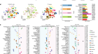
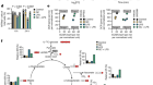
Medina, C. B. et al. Nature 580, 130–135 (2020).
- 2.
Green, D. R. Cell Death: Apoptosis and Other Means to an End 2nd edn (Cold Spring Harb. Lab. Press, 2018).
- 3.
Lüthi, A. U. & Martin, S. J. Cell Death Differ. 14, 641–650 (2007).
- 4.
Kerr, J. F. R., Wyllie, A. H. & Currie, A. R. Br. J. Cancer 26, 239–257 (1972).
- 5.
Henson, P. M. Annu. Rev. Cell Dev. Biol. 33, 127–144 (2017).
- 6.
Garg, A. D. & Agostinis, P. Immunol. Rev. 280, 126–148 (2017).
- 7.
Bosurgi, L., Hughes, L. D., Rothlin, C. V. & Ghosh, S. Immunol. Rev. 280, 8–25 (2017).
- 8.
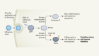
Chekeni, F. B. et al. Nature 467, 863–867 (2010).
- 9.
Haskó, G., Sitkovsky, M. V. & Szabó, C. Trends Pharmacol. Sci. 25, 152–157 (2004).
- 10.
Haskó, G. et al. J. Immunol. 164, 1013–1019 (2000).
- 11.
Orozco, S. L. et al. Cell Rep. 28, 2275–2287 (2019).
Competing Financial Interests
The author is a consultant for Inzen Pharmaceuticals and is on the scientific advisory board of Ventus Therapeutics.