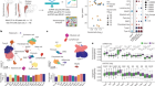
A researcher uses a confocal microscope equipped for super-resolution and fluorescence imaging at the University of Bristol, UK.Credit: Matt Lincoln/Wolfson Bioimaging/Univ. Bristol
Scientists are understandably wary of the word ‘revolution’. Major breakthroughs that shake the foundations of established thinking are rare compared with the small, additive steps by which science tends to progress. Yet when Werner Kühlbrandt at the Max Planck Institute of Biophysics in Frankfurt, Germany, wrote about the promise of a microscopic technique that could reveal the structure of large biomolecules at near-atomic resolution in 2014, he chose as his headline ‘The Resolution Revolution’1.
Kühlbrandt was referring to the potential of electron cryo-microscopy (cryo-EM), a technique that fires beams of electrons at proteins frozen in solution to reveal their structures. Cryo-EM is just one of many imaging tools driving rapid advances in cell biology and related fields. “New techniques have come on board over the last ten years that allow us to see things inside cells that we couldn’t resolve before,” says Anne Ridley, a cell biologist at the University of Bristol, UK, and president of the British Society for Cell Biology. “We can see whether two proteins are located in the same place, doing the same things, coming together or apart in real time, understand how enzymes work and obtain structures of complexes of proteins in ways we couldn’t have dreamed of doing before.”
Nature Career Guide: Cell biology
Scientists have been improving microscopes ever since the devices were invented in the late sixteenth century, and this process has accelerated markedly over the past decade. The 2014 Nobel Prize in Chemistry was awarded to the developers of super-resolution fluorescence microscopy, and the 2017 prize recognized the development of cryo-EM. The specifics of each microscopy technique vary hugely, as do the subsets of scientists that favour them. But, fundamentally, all are ways of seeing the cell in greater detail than is possible by eye.
Each microscope technique involves a compromise. Fluorescence microscopy, in which florescent molecules are used to light up target proteins, cells or cellular components, allows biologists to observe live samples in real time. But because visible light cannot distinguish between objects closer than 200 nanometres to each other, it is not, on its own, enough to reveal the detailed structures of tiny functional components in cells called organelles. Electron microscopes can achieve much higher resolutions, but require a vacuum and so cannot be used on live samples. In modern science, cell biologists have access to an ever-growing armoury of microscopy tools, which can be used either alone or in combination and offer many improvements over the much older technique of crystallography (see ‘Tools of the trade’).
Cross-disciplinary approach
These advances have been the result of contributions from scientists in many different fields. Physicists have provided much of the technology, such as the advanced electron detectors that increased the speed and sensitivity of modern cryo-EM devices. Chemists have developed brighter florescent probes that illuminate targets for longer. Statisticians and computer scientists have improved image processing and analysis techniques. “The acceleration in imaging has come about through this incredible synergy,” says Jennifer Lippincott-Schwartz, a cell biologist at the Howard Hughes Medical Institute’s Janelia Research Campus in Ashburn, Virginia, who helped to lay the foundations for the development of super-resolution microscopy with work during the 1990s on the use of green fluorescent proteins to visualize cellular trafficking pathways in living cells2.
Many advances have been made using these microscopy tools. Lippincott-Schwartz and her colleagues, for example, used a form of light-sheet fluorescence microscopy with confocal microscopy to capture 3D colour footage of the interactions between different types of organelle. “We were able to map out the relationships between six types of organelles, how fast they were moving and the contacts they made with each other,” says Lippincott-Schwartz, whose paper3 was published in Nature in 2017. “That’s important if you want to understand the cross-communication between organelles, which is one of the big interests among cell biologists right now.”

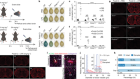
Jennifer Lippincott-Schwartz demonstrates a microscope capable of super-resolution imaging.Credit: Matt Staley
The growing availability of these advanced techniques presents opportunities for early-career cell biologists. Most obviously, it increases the number of processes cell biologists can probe. “These techniques open up tremendous vistas for the types of questions we can answer,” says Lippincott-Schwartz. Structural biologist David Barford at the MRC Laboratory of Molecular Biology in Cambridge, UK, has used cryo-EM to advance understanding of some of the cellular mechanisms involved in mitosis4, a type of cell division that results in the formation of two daughter cells with the same chromosomes as the parent cell. “For academic scientists, the ability to determine structures at atomic resolution with electron cryo-microscopy can be very important in the design of new experiments and testing of biological hypotheses,” he says.
Barford adds that the potential benefits to early-career researchers of acquiring an in-depth understanding of the latest imaging techniques could extend beyond the immediate research questions they are seeking to answer. “Drug companies are becoming very keen on electron cryo-microscopy as a means to determine the structures of proteins and drug targets, so moving into it could be a very good career choice,” he says. Barford also thinks these techniques will grow more important and overtake older techniques used by biologists. “It will probably supersede crystallography in the job market.”
It is impossible to become proficient in the use of all or even many of the latest imaging tools. Early-career cell biologists seeking to use them need to decide whether to specialize in a particular technique, or to identify collaborators who can do it for them (see ‘Meetings of minds’ for some of the conferences popular in cell biology). Ridley, who studies the role of cell migration in cancer progression, advises those doing PhDs to take up any opportunities available to them to get a flavour of the different techniques. “I would recommend that anyone doing a PhD programme with the option to do rotations in different labs and gain experience of different imaging areas to do so,” she says. “Even if you don’t become an expert in electron microscopy, for example, working in that area for a couple of months will give you an understanding of what it can and can’t do.” Barford adds that researchers who leave it to collaborators to do their imaging for them risk falling behind in other ways. “If you become just a user rather than a developer, it limits your future potential to contribute to the field through developing and advancing the technology.”
Meetings of minds

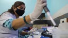
Delegates at the joint meeting of the American Society for Cell Biology and the European Molecular Biology Organization in 2018.Credit: Paul Sakuma Photography
Symposia and conferences are good for getting updates and overviews of a field.
Researchers often have to choose between going to broad or specialized meetings. For those seeking an overview of the state of the field, the joint meeting of the American Society for Cell Biology and the European Molecular Biology Organization is by far the largest annual gathering of cell biologists in the world. Around 6,000 people are expected to attend this year’s, in Washington DC on 7–11 December. Subjects to be covered will be wide-ranging, including emerging topics such as non-conventional model organisms, computational modelling and synthetic biology.
Bruce Stillman, president and chief executive of Cold Spring Harbor Laboratory in New York, will give the keynote lecture on his work on chromosome duplication in cells. There will be a variety of symposia, workshops, poster sessions and special-interest sessions. On the day before the main meeting, there will be a full day of session on careers and professional development for academics, and a one-day mini biotech course, at which attendees can learn how scientific discoveries are turned into bioscience ventures. Other sessions will cover careers in non-profit science advocacy, science policy, outreach, scientific infrastructure management and bench-based research in industry.
There are many other options for researchers wanting to dig deeper into a particular branch of the discipline. A symposium called Seeing is Believing, for example, brings together the developers of cutting-edge imaging techniques with those applying them in the lab. This meeting attracted some 400 participants when it was last held, at the European Molecular Biology Lab in Heidelberg, Germany, in October this year. It featured sessions on the latest tools and methods transforming researchers’ abilities to visualize proteins, protein complexes, organelles, cells, tissues, organs and whole organisms.
One of the draws of imaging for Lippincott-Schwartz is its purity as an empirical method for acquiring knowledge. “When you are imaging, you are first observing, then generating hypotheses and then designing approaches for testing your hypotheses. It’s the perfect avenue for fulfilling the scientific method.” She adds that the proliferation of advanced tools has made microscopy all the more attractive as a focus for cell biologists. “It can make imaging a very creative direction to take,” she says.
 A cell biology path outside of academia
 How a cell scientist in São Paulo is battling malaria
 The microscope makers putting ever-larger biological samples under the spotlight